Recognition and Management of Malnutrition
| Site: | Pediatric Pandemic Network Learn |
| Course: | Recognition and Management of Malnutrition |
| Book: | Recognition and Management of Malnutrition |
| Printed by: | Guest user |
| Date: | Saturday, September 12, 2026, 5:25 PM |
Table of contents
- 1. Overview
- 2. Introduction
- 3. Nutritional Status Assessment
- 4. Clinical Features of Malnutrition
- 5. Micronutrient Deficiencies
- 6. IMCI Strategy for Nutritional Status Assessment
- 7. Breastfeeding Programs in Disaster Situations
- 8. Nutritional Status of Infants 0 to 6 Months of Age
- 9. Summary
- 10. Suggested Reading
- 11. Case Resolution
- 12. Book Review
- 13. Appendix
1. Overview
Welcome to this multimedia open access edition of this chapter book titled Recognition and Management of Malnutrition Book.
This content is available as a digital book in two formats:
1) Website, which you are currently reading. This format offers a mobile friendly multimedia experience with sharable links to chapters. There are three ways to move through the book:
a) Use the table of contents chapter navigation to the left to navigate to different chapters in the book. Scroll through the links to select the chapter that interests you.
b) Use the yellow arrows that appear when scrolling to select previous and next chapters.

c) Use the previous chapter and next chapter buttons at the bottom of very page to navigate through the book.

2) Printable, which you can save as a PDF and open without an Internet connection.
1.1. Acknowledgements
Created by the Center for Global Health, Colorado School of Public Health
This content is owned by the Center for Global Health, Colorado School of Public Health, and has been jointly created by the Center for Global Health, Colorado School of Public Health and the Maternal and Child Health Department of Maimonides University; both of which are WHO Collaborating Centers in Maternal and Child Health. The course materials were developed with input from the American Academy of Pediatrics (AAP), the Pan American Health Organization (PAHO), and the Association for Health Research & Development (ACINDES).
With Support from the Pediatric Pandemic Network

The Pediatric Pandemic Network is supported in part by the Health Resources and Services Administration (HRSA) of the U.S. Department of Health and Human Services (HHS) as part of cooperative agreements U1IMC43532 and U1IMC45814 with 0 percent financed with nongovernmental sources. The content presented here is that of the authors and does not necessarily represent the official views of, nor an endorsement by HRSA, HHS, or the U.S. Government. For more information, visit HRSA.gov.

Attribution-NonCommercial-NoDerivatives CC BY-NC-ND
You are free to download and share this work for noncommercial purposes, as long as you credit the Center for Global Health and the Pediatric Pandemic Network for the original creation.
2. Introduction
Book Authors
Daniel Martinez-Garcia MD | Laurent Hiffler,MD | Teresa M. Kemmer, PhD, RD | Clifton Yu, MD | Andrew J. Bauer, MD | Julia A. Lynch, MD
Introduction
Providing adequate food to meet the nutritional needs of growing children is critical to prevent an increase in malnutrition prevalence, which would lead to excess mortality during the recovery phase of a disaster. Nutritional status directly impacts the vulnerability for and the severity of infectious diseases that affect children in emergency settings. Children having frequent infections have associated anorexia, which increases the stress burden and severity of malnutrition. Previously malnourished children are particularly vulnerable, as they cannot develop the protective compensatory mechanisms that allow healthy individuals to survive during periods of food deprivation. Therefore, after disasters those children will decompensate unless given immediate nutritional support. On the other hand, good nutritional status promotes wound healing and improves the postnatal outcomes in both mothers and babies. In disaster situations, it is crucial to provide adequate food to prevent the complications associated with malnutrition. According to the World Health Organization (WHO), “food security exists when all people, at all times, have access to sufficient, safe and nutritious food to meet their dietary needs and food preferences, and to maintain an active and healthy life.”
Doctors and nurses and nutrition specialists and epidemiologists in the local community are an invaluable source of information regarding the predisaster nutritional status of the pediatric population. Their involvement in nutritional assessments and food resource planning is essential. During an assessment, recognize that large variations in baseline pediatric nutritional status can be found in a particular region. In addition, a paradoxical association of malnutrition and obesity can be found within the same household, especially in developing countries and in low-income groups. Micronutrient deficiencies, most importantly iron deficiency anemia, can be identified even in areas where the overall nutritional status is good.
3. Nutritional Status Assessment
OBJECTIVES
- Recognize the importance of assessing the nutritional status of the pediatric population affected by a disaster.
- Identify the vulnerable population groups and specific risk factors in these situations.
- Know and use the different methods for anthropometric assessment of the pediatric population.
Initial Assessment
The initial assessment of the nutritional status and food resources of a population affected by a disaster is part of the overall emergency needs assessment (Box 1). Obtain all available information regarding the pre-disaster prevalence of macro- and micronutrient deficiencies in the community. Information from public health authorities, health care professionals, and other health workers in the local community is critical for that purpose. Also, identify any nutrition programs active in the community before the disaster. This information helps to identify nutritionally vulnerable groups (Box 2). Finally, determine the quantity and quality of food stores readily available to the affected population. The data obtained through the initial assessment, together with accurate demographic data from the affected population, are needed to design and implement an adequate food response following a disaster.
BOX 1. Assessment of the nutritional status and resources of the population affected by a disaster
- Initial assessment
- Determine malnutrition and micronutrient deficiency prevalences before the disaster
- Identify nutrition programs active in the community before the disaster
- Identify nutritionally vulnerable groups
- Determine the quantity and quality of available food storages readily available to the affected population
- Determine social, cultural, economical and political determinants that could impair the fair distribution of food resources among the affected population.
- Assessment during the recovery phase
- Determine the quality and security of available nutritional resources for the affected population, particularly for vulnerable groups
- Determine current prevalence of malnutrition and micronutrient deficiencies
- Do periodic reassessments until adequate nutrition resources are sustainable.
BOX 2. Vulnerable groups in a disaster situation
- Children under 5 years of age
- Children/adolescents taken away from their family or community or have lost a parent
- Pregnant or lactating women
- Families living in a household headed by a woman
- Physically or emotionally disabled persons
- People with chronic diseases
- Elderly people
- Families having lost their home or job as a direct consequence of the disaster
Assessment during the recovery phase
In the recovery phase, as more outside resources become available and the local community becomes more organized, one of the goals is the development of programs to guarantee that available food resources will be targeted efficiently and effectively to populations in need. This requires the systematic assessment of the nutritional status of the population. Box 1 outlines the basic components of ongoing recovery phase nutritional assessments. These measures should be continued until adequate nutrition resources are appropriately and efficiently distributed.
Anthropometric assessment in the pediatric population
Anthropometric methods provide information regarding the height, weight, and proportions of a person. These data are used, particularly in children, to assess an individual’s nutritional status. Interpretation of anthropometric data requires the comparison of the individual’s measurements to standards for the appropriate population. When anthropometrics are systematically collected in a population, it is possible to characterize the community’s overall nutritional status. Generally the data from children under 5 years of age reflect the status of the community. In disaster situations, such data help to determine the global nutritional needs for all the affected population and how resources should be efficiently allocated.
Case 1
You are member of a medical team delivering health care in a small town that has suffered a serious flood. It is necessary to establish the nutritional needs of the affected population.
- What are the first steps in such evaluation?
The assessment of the nutritional status of a population affected by a disaster is an ongoing process. It begins during the rescue phase with an initial rapid assessment and should continue as attempts are made to efficiently and equitably provide adequate nutrition resources to the affected population. The information gathered through this assessment is needed to use available resources more rationally during both the initial and the recovery phases of a disaster.
Anthropometric indexes
Box 3 shows the anthropometric indexes most commonly used in the assessment of children.
BOX 3. Anthropometric indexes most frequently used in children
- Weight-for-age
- Weight-for-height
- Height-for-age
- Mid-upper arm circumference
- Body mass index
Weight-for-age index (W/A)
The W/A index represents the weight of a child in relation to his or her age. Consider the presence of dehydration and edema, which alter the weight when determining the index. A precision scale is required for weight measurement.
Weight-for-height index (W/H)
The W/H index represents the weight of a child in relation to the height. It reflects the current nutritional status of the child and is the index used to diagnose acute (wasting) or subacute malnutrition. It also requires a precision scale and a measuring board or tape, which are not usually readily available in disaster situations and even if available take considerable time to obtain. W/H is also affected by dehydration and edema.
Anthropometric methods provide information regarding the height, weight, and proportions of a person. These data are used, particularly in children, to assess the nutritional status of an individual.
Height-for-age (H/A) index
The H/A index represents the height of a child in relation to his or her age. This index basically reflects the nutritional history, since children with chronic malnutrition— whether primary or secondary to an underlying chronic disease—will experience stunted growth. Height, however, is also strongly determined by genetic factors as well as mothers nutritional status.
Mid-upper arm circumference (MUAC)
The MUAC gives a measure of the amount of fat and muscle in the upper arm. It is measured with a standard tape on the left arm, midpoint between the shoulder and the tip of the elbow. It is used in children 6 months to 5 years of age to screen large numbers of children for malnutrition. For children from 10-18 years of age, MUAC is an unreliable method to identify malnutrition. For adults above 18 years of age, an MUAC cut off of less than 185 mm indicates severe acute malnutrition.
Body mass index (BMI)
BMI is the weight in kilograms divided by the height in meters, squared (weight in kg/[height in m]2). BMI reference tables are now available to be used in children and adolescents from 2 to 20 years old. As an index, BMI does not reflect small changes in weight that may be clinically relevant, and it is also affected by dehydration and edema. Cut-off values for BMI percentiles in children and adolescents are as follows:
BMI Percentile Classification
| BMI percentile | Classification |
|---|---|
| <5th | Underweight |
| 5th - 85th | Within normal limits |
| 85th - 95th | At risk for overweight |
| >95th | Overweight |
Percentiles
Percentiles are determined by the position of an individual’s measure in the reference values in terms of the percentage of values exceeded or equaled. In the reference population, the weight for a given height shows a normal distribution. The 50th percentile is the weight that divides the reference population into two equal parts: with 50% above and 50% below. As an example, if 25% of the reference population weighs less than the child being examined, the child is in the 25th percentile.
A review of how these anthropometric tools may be used to determine nutritional status of an individual is shown in Table 1.
TABLE 1. Assessment of nutritional status with anthropometric indexes
Nutritional Status Classification
| Nutritional Status | MUAC Age 6-59 months | W/H% | W/H Z-score | Percentile |
|---|---|---|---|---|
| Moderate acute malnutrition | 115–125 mm | >70 and <80% | -2 to -3 SD | |
| Severe acute malnutrition | <115 mm | <70% or edema | < -3 SD or edema | |
| Underweight | BMI <5th |
Reference tables
Regardless of the anthropometric parameter used, the measurements obtained are useful only if the standards with which they are compared truly reflect the population that is being evaluated. Many countries have developed their own growth tables and graphics reflecting the standard for their own population, but many regions have not been included. WHO recently published new growth reference charts developed with data gathered from Brazil, Ghana, India, Norway, Oman, and the United States. The children selected were exclusively breastfed, healthy, and had their basic needs met. There is solid evidence to suggest that all children up to age 5 years grow very similarly when their physiologic needs are adequately met, so these reference charts are valid for evaluating growth in children all over the world. More information can be found at http:/www.who.int/childgrowth/en/.
4. Clinical Features of Malnutrition
Objectives
- Identify through physical examination the main clinical findings of protein-energy malnutrition and those indicating severe malnutrition.
- Recognize the particular features and the clinical and pathophysiologic differences between marasmus and kwashiorkor.
- Describe the pathophysiology of the refeeding syndrome.
Healthy, well-nourished persons have some protection from acute malnutrition, because they have adequate stores of glycogen, protein reserves, and calories stored as fat. During the first 3 days without food, glycogen stores in the liver and muscle are depleted, and the liver attempts to maintain blood sugar levels by converting mobilized amino acids into glucose (gluconeogenesis). At the same time, the breakdown of fat (lipolysis) leads to the formation of an alternate fuel source, ketone bodies, allowing for short-term survival. However, individuals who are malnourished at the onset of a disaster are incapable of activating these protective mechanisms and thus face greater risk of acute nutritional decompensation.
The pathophysiology of severe protein-energy malnutrition is very complex, affecting the cellular function of many organ systems, including heart and bowel. In the heart, redistribution of muscle proteins increases the risk for cardiovascular collapse, and damage to the intestinal villi leads to malabsorption.
Marasmus is the most common form of PEM and is caused by deprivation of both energy/calories and protein that leads to weight loss of more that 20% of initial body weight. It is characterized by profound wasting, fatigue, apathy, and irritability.
4.1. Types of protein-energy malnutrition
Protein-energy malnutrition (PEM) is a general term describing a state of deficiency involving multiple nutrients. Usually malnutrition in children results from a combination of energy and protein deficiency, often with associated micronutrient deficiency. Frequent infections causing anorexia and decreased food intake play an important contributing role. The pathophysiology of severe PEM is very complex, affecting the cellular function of many organ systems, including heart and bowel. In the heart, redistribution of muscle proteins increases the risk for cardiovascular collapse, and damage to the intestinal villi leads to malabsorption. The edema associated with malnutrition results from a combination of hypoalbuminemia and deficiencies in copper, zinc, selenium, thiamine and vitamins A, E, and C.
There are two major clinical presentations of severe PEM: marasmus and kwashiorkor. Marasmus is the most common form of PEM and is caused by deprivation of both energy/calories and protein that leads to weight loss of more that 20% of initial body weight. It is characterized by profound wasting, fatigue, apathy, and irritability. A person of normal weight (10% to 12% body fat) would develop marasmus after approximately 60 days of total starvation. Marasmus is most common in infants under 1 year of age, and these children maintain their hunger despite appearing irritable.
Kwashiorkor usually occurs when a previously malnourished patient is exposed to the catabolic stress of infection (measles, tuberculosis, pertussis, and others), diarrhea, or trauma. Studies suggest that there is no difference in diets of children who develop marasmus or kwashiorkor. The generation of free radicals and depletion of anti-oxidants associated with inflammation appears to be linked to the development of edema in kwashiorkor. Nutritional edema is associated with an increased secretion of an anti-diuretic substance (probably antidiuretic hormone) which prevents the normal excretion of free water. Low-protein, low-calorie diets may affect the inactivation of anti-diuretic hormone.
In Latin America, kwashiorkor is estimated to occur in only 2% of malnourished individuals, although in some regions in the world, such as Africa, this proportion is much higher, up to 30%. It is more commonly found in children 1 to 3 years of age and is more prevalent in regions where the majority of nutrition is obtained from starchy vegetables that may be contaminated with aflatoxin (a fungal toxin that commonly attacks plants in wet regions). Kwashiorkor is characterized by abdominal distension, peripheral edema, flaking skin lesions, hair changes, including decoloration, and hepatomegaly. Children with kwashiorkor are often anorexic, which poses additional challenges to their management. A subset of these children may present with marasmic kwashiorkor, typically with edema, significant subcutaneous fat and muscle wasting, stunting, and mild hepatomegaly. Children with marasmic kwashiorkor show high mortality rates; thus, highly cautious rehydration and refeeding are critical.
Kwashiorkor is characterized by abdominal distension, peripheral edema, flaking skin lesions, hair changes, including decoloration, and hepatomegaly. Children with kwashiorkor are often anorexic, which poses additional challenges to their management.
4.2. The Refeeding Syndrome
Complications of refeeding syndrome include:
- Hypomagnesemia
- Hypoglycemia
- Hypokalemia
- Hypophosphatemia
- Thiamine deficiency
During starvation, there is a redistribution of proteins, fluids, and electrolytes, as the body tries to adapt to the state of malnutrition. Acidosis, associated with the catabolic state, leads to a potassium shift from the intracellular compartment into the blood. Elevated aldosterone levels result in total body potassium depletion (similar to that found in diabetic ketoacidosis). Reintroduction of fluids and carbohydrates, via oral, enteral, or parenteral routes, produces a sudden shift back to glucose as the predominant fuel source, leading to increased demand for phosphorylated intermediates of glucose metabolism, increased insulin production, and the shift of potassium back into the cells associated with the resolution of the acidosis. This results in hypokalemia and hypomagnesemia. Acute thiamine deficiency syndrome shares many common elements with the refeeding syndrome but is a separate entity. It is also very frequent in children with Severe Acute Malnutrition. The refeeding process with carbohydrate drives a rapid use of Thiamine that produces a “functional Thiamine deficiency” aggravated by low thiamine body stores.
New studies of cardiac function in children with severe acute malnutrition do not support the long held belief that these children have poor function and will not tolerate a high volume of fluids. Insulin may play a key role in the development of pulmonary edema and congestive heart failure in these children by exerting an anti-diuretic effect leading to sodium and water retention that result in the expansion of the extracellular water compartment.
Therefore, it is necessary to reassess the patient repeatedly and develop an appropriate management plan to avoid these complications. Give additional phosphate, potassium, magnesium, and thiamine, as well as a continuous supply of glucose to compensate for rapid shifts between intracellular and extracellular compartments (see Appendix for suggested recommendations). When refeeding is initiated, it should be performed in phases (see Section IV). Consider treating associated infections and likely micronutrient deficiencies.
5. Micronutrient Deficiencies
Objectives
- List specific micronutrient deficiencies, their risk factors, and clinical signs.
- Describe the epidemiology, pathophysiology, and clinical presentation of vitamin A, iron, and zinc deficiencies.
- Describe the general management for micronutrient deficiency prevention and treatment in acute emergency settings.
Different dietary insufficiencies may lead to specific micronutrient deficiencies. Some of these have typical clinical manifestations. Specific dietary risk factors for important micronutrient deficiencies and possible solutions are outlined in Table 2. Table 3 summarizes the classic physical findings associated with significant micronutrient deficiencies.
Case 2.
You are providing pediatric care to a population that has been displaced after a flood. A mother comes to the health care unit with her 4-month old son, who was born after 35 weeks of gestation and is exclusively breastfed. Physical findings are normal. The weight and height are at the 50th percentile (age corrected for gestational age).
- What are your recommendations regarding iron intake?
TABLE 2. Micronutrient deficiencies: risk factors and possible solutions
| Niacin (pellagra) | |
|---|---|
| Dietary risk factor | Maize-based diet |
| Possible solutions | Foods rich in proteins and whole grain cereals |
| Thiamin (beri-beri) | |
|---|---|
| Dietary risk factor | Polished rice/or other cereal/cassava/manioc-based diet |
| Possible solutions | Whole or parboiled rice, legumes, beef, fish, eggs, milk; fortified cereal blends |
| Vitamin A | |
|---|---|
| Dietary risk factor | Diet with not enough fresh fruits |
| Possible solutions | Dark orange fruits and vegetables, yellow corn, fortified cereal, animal products, dark green leafy vegetables, amaranthus, baobab, mango leaves, vitamin A supplements |
| Vitamin C (scurvy) | |
|---|---|
| Dietary risk factor | Diet with not enough fresh fruits and extremely low fat intake |
| Possible solutions | Fresh raw fruits/vegetables, liver, fresh animal milk |
| Iron (ferropenic anemia) | |
|---|---|
| Dietary risk factor | Diet lacking animal products |
| Possible solutions | Animal products (liver, meat); dried fruits; consumption of vitamin C with meals; iron/folate supplements or fortified cereal blends. From ages 6-24 months on, nearly all iron intake is provided by supplementary foods. |
| Zinc | |
|---|---|
| Dietary risk factor | Diet lacking animal products |
| Possible solutions | Animal products (liver, meat); fortified cereal, peanuts, Gombo (Okra), sesame and pumpkin seeds, avocado, all legumes: niebe, lentils, peas, Moringa leaves powder. Children 6-24 months on, millet, sorgho. From ages 6-24 months on, nearly all zinc intake is provided by supplementary foods. |
| Riboflavin | |
|---|---|
| Dietary risk factor | Diet lacking animal products |
| Possible solutions | Animal products (liver, eggs, fish), milk, leafy green vegetables. From ages 6-24 months on, nearly all riboflavin intake is provided by supplementary foods. |
| Vitamin D (rickets) | |
|---|---|
| Dietary risk factor | Lack of exposure to sunlight |
| Possible solutions | Apart from a supplement diet vitamin D is negligible |
| Calcium | |
|---|---|
| Dietary risk factor | Lack of milk, dark-green leaves, or fish with bones |
| Possible solutions | Milk, fish with bones (e.g. sardines), fish flour, shell fish, egg shell powder, beans and green peas, dark green leaves, calcium carbonate (used in making tortillas) |
TABLE 3. Physical findings commonly associated with micronutrient deficiencies
| Hair | |
|---|---|
| Physical signs | Dry, dull, easily pluckable; sparse hair loss |
| Possible nutritional deficiencies | Protein-energy malnutrition, zinc, protein, biotin, essential fatty acids |
| Eyes | |
|---|---|
| Physical signs | Pale sclera; Bitot’s spots; night blindness |
| Possible nutritional deficiencies | Iron, vitamin B6, B12, vitamin A |
| Mouth | |
|---|---|
| Physical signs | Red swollen lips; Angular stomatitis (cracks at sides of mouth); Cheilosis |
| Possible nutritional deficiencies | Niacin, riboflavin, iron and/or vitamin B6; Niacin, riboflavin, iron and/or vitamin B6; Niacin, riboflavin |
| Gums | |
|---|---|
| Physical signs | Swollen, bleeding, abnormally red |
| Possible nutritional deficiencies | Vitamin C |
| Tongue | |
|---|---|
| Physical signs | Glossitis; Dark red; Pale |
| Possible nutritional deficiencies | Vitamin B complex*; iron; riboflavin |
| Teeth | |
|---|---|
| Physical signs | Dental caries |
| Possible nutritional deficiencies | Fluoride, vitamin C |
| Taste | |
|---|---|
| Physical signs | Dysgeusia or hypogeusia |
| Possible nutritional deficiencies | Zinc |
| Skin | |
|---|---|
| Physical signs | Loose; Lower extremity edema; Pallor; Poor healing; Rough skin, poor turgor (positive skin pinch); Small, purplish spots; Pellagra (pigmented keratotic scaling lesions); Follicular hyperkeratosis; Ecchymosis |
| Possible nutritional deficiencies | Calories; Protein, thiamine; Folic acid, thiamine, vitamin B12, biotin; Vitamin C; Fluids, marasmus, protein, calories; Vitamin C; Niacin; Vitamin A and/or essential fatty acids; Vitamin K |
| Nails | |
|---|---|
| Physical signs | Brittle or ridged spoon-shaped |
| Possible nutritional deficiencies | Protein; Iron |
| Musculoskeletal | |
|---|---|
| Physical signs | Muscle wasting; Rickets, osteomalacia |
| Possible nutritional deficiencies | Protein, energy; Vitamin D, phosphorus, calcium |
| Neurological | |
|---|---|
| Physical signs | Hyperreflexia; Ataxia; Encephalopathy; Nystagmus; Peripheral neuropathy |
| Possible nutritional deficiencies | Vitamin B6; Thiamine; Sodium chloride |
5.1. Vitamin A Deficiency
Vitamin A is critical for vision and epithelial integrity. In addition, vitamin A deficiency (VAD) is associated with disorders in hematopoiesis and immune function. Thus, treatment of such deficiency has beneficial effects for patients with anemia and improves the outcome of infections, particularly measles. VAD is associated with diets lacking fresh fruits and vegetables, as well as animal products, dairy products, and eggs. VAD has a dramatic global impact on health, with approximately 127 million preschool-age children and 20 million women affected worldwide. It has been estimated that unidentified VAD results in about 2 million deaths in young infants, particularly due to excess morbidity and mortality associated with measles (see Module 5). VAD is the most common preventable cause of childhood blindness in the world. It is also the most frequent deficiency syndrome among displaced populations.
The clinical features of VAD involving the ocular system are known as xerophthalmia. The stages of xerophthalmia include night blindness, conjunctival xerosis, and keratomalacia. Night blindness is the most prevalent and earliest stage of xerophthalmia resulting from the impact of VAD on the retinal epithelium. Since this symptom may precede any apparent physical findings, its occurrence must be assessed through a careful history. Conjunctival xerosis presents as a dry nonwettable, rough or granular surface, which can be seen using a hand-light. More advanced xerosis is associated with Bitot’s spots which are bubbly, foamy, or cheese-like patches visible on the conjunctival epithelium. Conjunctival xerosis may progress to ulceration or in the most advanced form to keratomalacia, its typical presentation being necrosis of the cornea.
It has been estimated that unidentified vitamin A deficiency results in about 2 million deaths in young infants, particularly due to excess morbidity and mortality associated with measles.
Supplementation
A diet containing sufficient amounts of foods rich in vitamin A is enough to prevent hypovitaminosis. When adequate amounts of vitamin A are not available through dietary sources, consider supplementation. Vitamin A supplementation has been shown to reduce pre-school child mortality by 25% to 35%, and to virtually eliminate nutritional blindness in many low- and middle-income countries.
In acute humanitarian emergencies, if an adequate diet was not available and a regular vitamin A supplementation program was not in place for the general population prior to the disaster, provide vitamin A supplementation to all children 6 months to 5 years of age at the first contact with the health care staff. Remember to check whether the child already received vitamin A as part of any mass vaccination campaign. Fortified foods with vitamin A and other essential micronutrients should be distributed during the recovery phase. Individuals with symptoms and signs of VAD should receive the recommended treatment. Table 4 shows preventive and treatment doses of vitamin A. Only provide preventive treatment with vitamin A when it is known that the population is deficient.
| <6 months (<6 kg) | |
|---|---|
| Treatment | 50,000 IU |
| Preventive dosage | 50,000 IU every 4-6 months |
| 6-12 months (6-8 kg) | |
|---|---|
| Treatment | 100,000 IU |
| Preventive dosage | 100,000 IU every 4-6 months |
| >1 year (>8 kg) | |
|---|---|
| Treatment | 200,000 IU |
| Preventive dosage | 200,000 IU every 4-6 months |
| Women | |
|---|---|
| Treatment | 200,000 IU** |
| Preventive dosage | 200,000 IU ≤ 8 weeks after delivery |
Notes:
- * Treat all cases of xerophthalmia and measles with the same age-specific dosage the next day and again 1 to 4 weeks later.
- ** For women of reproductive age, give 200,000 IU only for corneal xerophthalmia; for milder eye signs (night blindness or Bitot’s spots), give 5,000-10,000 IU per day or ≤25,000 IU per week for ≥4 weeks.
5.2. Iron Deficiency
Iron deficiency (ID) is the most common nutritional deficiency worldwide. In developing countries, most affected individuals are women and children. Risk factors for ID, in addition to a diet lacking animal products, include: pregnancy, prematurity, low birthweight, early umbilical cord clamping, rapid growth, cow’s milk feeding (intestinal microhemorrhages), reduced intestinal absorption of iron due to high phytate and phosphate intake (cola beverages), menstruation, and parasitic infections. ID is also the most frequent cause of anemia. The three major causes of anemia in the developing world are nutritional deficiencies, malaria, and intestinal parasites (hookworm). The prevalence of anemia has been used as a surrogate marker of ID prevalence in a certain population. It has been estimated that ID in a population is 2 to 3 times more prevalent than ID anemia (IDA).
Clinical findings associated with severe anemia include skin, mucus membranes, and nail beds pallor, as well as dyspnea or tachypnea at rest. Clinical examination is not a reliable method for diagnosing isolated iron deficiency or milder forms of anemia. If laboratory tests are available, the diagnosis of anemia can be documented with hemoglobin (Hb) or hematocrit determinations. Table 5 shows the age-specific cut-off values for Hb and hematocrit according to WHO guidelines. The reduction in tissue oxygen supply associated with anemia is responsible for the clinical manifestations and long-term consequences of iron deficiency. Anemia is associated with growth retardation, increased susceptibility to infections, and impaired cognitive and psychomotor development. Very severe anemia (Hb <5 g/mL) is associated with increased mortality. Iron therapy and multivitamin therapy have both been shown to reverse some of these effects, but long-term studies suggest that iron deficiency anemia in early childhood can lead to irreversible developmental damage.
TABLE 5. Hemoglobin (Hb) and hematocrit (Hct) cut-off values used to define anemia in people living at sea level
| Age or Group | Hb below | Htc below |
|---|---|---|
| Children 6 to 60 months | 11 g/mL | 33% |
| Children 5-11 years | 11.5 g/mL | 34% |
| Children 12-13 years | 12 g/mL | 36% |
| Non-pregnant women | 12 g/mL | 36% |
| Pregnant women | 11 g/mL | 33% |
| Men | 13 g/mL | 39% |
** Iron deficiency is the most common nutritional deficiency worldwide.**
Iron supplementation for prevention and treatment of anemia
Due to the high bioavailability (about 50%) of lactoferrin-linked iron in human milk, exclusive breastfeeding during the first 4 to 6 months guarantees an appropriate iron pool in healthy term infants. Preterm infants need early iron supplementation, because their iron pools at birth are insufficient. With the introduction of solids at 6 months of age, begin appropriate supplementary feeding including foods with highly bioavailable heme iron (see Table 8 on page 19). Iron absorption can be enhanced by adding animal protein to the food.
Adequate intake of vitamin C and reduction of iron absorption suppresors in the diet also help to increase iron bioavailability. Adequate dietary intake of folic acid is also important, since IDA is often associated with folate deficiency (see Table 8 on page 19).
Due to the high bioavailability (about 50%) of lactoferrinlinked iron in human milk, exclusive breastfeeding for 4-6 months guarantees adequate iron pools in healthy full-term infants.
Iron supplementation programs have been effective in preventing ID. Preventive iron supplementation beginning at 6 months of age is encouraged and should be made available to those at risk for ID, since risks associated with ID at this age are highly significant. Recommendations for iron combined with folic acid supplementation for the prevention of ID are found in Table 6; severe anemia management is outlined in Table 7.
TABLE 6. Iron supplements to prevent anemia
| Children 6-12 months - Prevalence <40% | |
|---|---|
| Dosage (daily) | 12.5 mg iron plus 50 μg folic acid |
| Duration | From 6-12 months of age |
| Children 6-12 months - Prevalence >40% | |
|---|---|
| Dosage (daily) | 12.5 mg iron plus 50 μg folic acid |
| Duration | From 6-24 months of age |
| Children 2-5 years | |
|---|---|
| Dosage (daily) | 20-30 mg iron plus 50-150 μg folic acid |
| Duration | |
| Children 6-11 years | |
|---|---|
| Dosage (daily) | 30-60 mg iron plus 50-150 μg folic acid |
| Duration | |
| Adolescents and adults | |
|---|---|
| Dosage (daily) | 60 mg iron (girls and women of reproductive age should also receive 400 μg folic acid) |
| Duration | 2-4 month course of daily dosing or weekly supplementation for as long as they are at risk |
| Pregnant women - Prevalence <40% | |
|---|---|
| Dosage (daily) | 60 mg iron + 400 μg folic acidᵃ |
| Duration | Six months in pregnancy (or if started late, extend to postnatal period for a total of 6 months)ᵇ |
| Pregnant women - Prevalence >40% | |
|---|---|
| Dosage (daily) | 60 mg iron + 400 μg folic acidᵃ |
| Duration | Six months in pregnancy plus continuing to three months post-partum (or a total of 9 months) |
Notes:
- ᵃ Folic acid supplementation is included with iron supplementation.
- ᵇ If supplementation begins late during pregnancy, continue into the postnatal period to complete the total recommended duration.
TABLE 7. Treatment for severe anemia
| <2 years | |
|---|---|
| Dosage (daily) | 25 mg iron plus 100-400 μg folic acid |
| Duration | 3 months |
| 2-12 years | |
|---|---|
| Dosage (daily) | 60 mg iron plus 400 μg folic acid |
| Duration | 3 months |
| Adolescents | |
|---|---|
| Dosage (daily) | 120 mg iron plus 400 μg folic acid |
| Duration | 3 months |
| Adults | |
|---|---|
| Dosage (daily) | 60-120 mg iron |
| Additional supplementation | Plus 1,000 μg folic acid |
| Duration | 2 months (iron), 15-30 days (folic acid) |
Note: Iron dosage for children 2-5 years of age is based on 2 mg iron/kg body weight/day Adapted from Nutrition Essentials: A Guide for Health Managers (2004). BASICS II.
Public Health Measures: To ensure adequate iron status, in addition to providing adequate amounts of dietary iron, it is essential to implement public health programs to control hookworm and other micronutrient deficiencies. In selected areas with endemic infections, antihelminthic medication should also be routinely given to all individuals over the age of 2 years, since helminthic infections, including hookworm infections, can have significant negative impact on anemia status. Various medications are available for the treatment of helminthic infections. For example: mebendazole (100 mg tablets) is used for routine therapy in hookworm infection in adults and children over 2 years: 1 tablet morning and evening for 3 days. For global therapy in a community, a single 200 mg dose is provided to all individuals over 2 years.
5.3. Zinc deficiency
The exact prevalence of zinc deficiency worldwide is not known, but is estimated to be similar to that of ID, which makes it an underrecognized public health problem. Zinc is essential for mammalian cell life, function, growth, differentiation, and replication. Yet it is one of the least apparent micronutrient deficiencies. Zinc plays a central role in protecting health and immune function of individuals, as a constituent of more than 200 enzymes and transcription proteins that modulate cell differentiation, nucleic acid synthesis, and the metabolism of proteins, lipids, and carbohydrates.
Zinc supplementation in children with deficiency has been shown to reduce the incidence and prevalence of diarrhea and severe lower respiratory tract infections. Supplementation with zinc also reduces the frequency of malaria infections.
Decreased growth velocity or stunted growth is a consistent and early outcome of even mild zinc deficiency in infants, children, and adolescents. Box 4 shows the multiple clinical features of zinc deficiency.
BOX 4. Clinical manifestations of zinc deficiency
- Decreased growth velocity
- Peri-orificial and acral skin lesions
- Glossitis
- Alopecia
- Nail dystrophy
- Dry inflamed scaly skin
- Delayed sexual development
- Erectile dysfunction
- Behavioral abnormalities
- Depressed mood
- Photophobia and impaired ocular adaptation to darkness
- Delayed wound, burn, and ulcer healing
- Impaired or loss of taste
- Low birth weight and prematurity
Risk factors for zinc deficiency include: insufficient dietary intake (low-protein diet); high phytate and/or fiber content in the diet; diarrhea and other malabsorption syndromes; intestinal parasitosis; hot and humid weather; and no breastfeeding.
Young children’s dietary intake of zinc appears to be inadequate in many developing countries and it has been estimated that 80% of women globally and 100% of women in developing countries, have zinc intakes inadequate to meet pregnancy needs. Food sources high in zinc are listed in Table 8 (page 19).
TABLE 8. Food sources high in selected vitamins and minerals
| Vitamin A | |
|---|---|
| Food sources, absorption inhibitors, and enhancers | Plant foods high in vitamin A (carotenoids): Greens (spinach, chicory, endive, collard, watercress, mustard, beet, turnip, broccoli), leaves of Amarantus, Manioc/cassava leaves, Baobab leaves, carrot, pumpkin, orange flesh sweet potato (the other form is not so rich in vit A), squash (winter acorn, hubbard and butternut), peas, red hot chili and sweet peppers, mango, papaya, apricot, asparagus, tomato, prune, plum. Preformed vitamin A from animal foods is found in mother’s milk, liver, fish-liver oils, butter, cheese, milk fat, eggs and vitamin A-fortified foods. |
| Iron | |
|---|---|
| Food sources, absorption inhibitors, and enhancers | Heme iron: Meat, fish, and poultry Non-heme iron: Eggs, dried beans, green leafy vegetables, whole grains, legumes, seeds, dried fruits, cumin seeds, anise seeds, Néré (sumbala a traditionally fermented preparation used in sub-saharan africa with Nere seeds), african snail meat, curry, molase (do not encourage refined sugar), pain de singe (baobab fruit also rich in vit C), dried ants and iron-fortified foods Absorption enhancers: Foods containing Vitamin C, other organic acids, and animal tissue Absorption inhibitors: High phytate foods, such as maize, legumes, whole wheat, brown rice and unmilled sorghum. Foods high in tannins (polyphenol) such as tea and coffee |
| Folic acid | |
|---|---|
| Food sources, absorption inhibitors, and enhancers | Green leafy vegetables, such as spinach and romaine lettuce; pinto, kidney, and navy beans; peas; chicken giblets; liver; strawberries; citrus fruits and juices; peanut; whole grain breads, rolls, crackers, and cereals; and fortified cereals, pasta, rice, and flours |
| Niacin and tryptophan | |
|---|---|
| Food sources, absorption inhibitors, and enhancers | Niacin: Meat, poultry, fish, liver, peanuts (groundnuts), legumes, and yeast Increases the bioavailability of niacin: Alkali processing Tryptophan (metabolizes into niacin): Milk and eggs |
| Thiamine | |
|---|---|
| Food sources, absorption inhibitors, and enhancers | Thiamine content is very high in fish such as trout, salmon, tuna. Also in seeds such as sunflower seed. Also squash, soy beans and beans. Parboiled rice, whole grain flour and cereals, pulses, nuts, wheat germ, yeast extract, pork, liver, kidney, and vegetables, such as peas, asparagus, and okra Absorption enhancers: Foods containing vitamin C Absorption inhibitors: Tea, coffee, alcohol and folate deficiency, betle nut and Thiaminase in some fish and larvae. Also avoid rinsing rice too many times. |
| Vitamin C (ascorbic acid) | |
|---|---|
| Food sources, absorption inhibitors, and enhancers | All citrus juices and fruits, such as orange, lemon, lime, kiwi, guava, and grapefruit; cabbage; tomato; berries; potatoes with skins; green and red peppers; broccoli; spinach; and brussels sprouts |
| Vitamin D | |
|---|---|
| Food sources, absorption inhibitors, and enhancers | Dairy products, fortified milk, fortified cereals, eggs, oily fish, such as herring, salmon, or tuna, and fish liver oils. Try to ensure adequate sun exposure. |
| Iodine | |
|---|---|
| Food sources, absorption inhibitors, and enhancers | Fortified foods, such as iodized salt are required in areas of the world that have an inadequate amount of iodine in the soil |
| Zinc | |
|---|---|
| Food sources, absorption inhibitors, and enhancers | Red meat, liver and other viscera, poultry, lamb, shellfish, eggs, and milk are excellent sources of bioavailable zinc. Peanuts, peanut butter, legumes, okra, sesame, Amarantus leaves, moringa leaves, pumpkin leaves, avocado, unpolished cereals |
| Vitamin B6 (pyridoxine) | |
|---|---|
| Food sources, absorption inhibitors, and enhancers | Milk, whole grain cereals, bread, liver, avocados, spinach, green beans, banana, fish, poultry, meat, nuts, potatoes, green leafy vegetables |
Promotion of exclusive breastfeeding for the first 6 months prevents zinc deficiency in infants. Fruits and other vegetables are not good sources of zinc, because zinc in vegetable proteins is poorly bioavailable, in contrast to zinc associated with animal proteins. It is also important to reduce the phytic acid content of the diet because it suppresses zinc absorption.
Supplementation offers the most immediate approach to improving zinc status, and fortification should be the primary long-term public health initiative to prevent deficiencies of this micronutrient. Box 5 shows zinc daily recommended intakes.
BOX 5. Zinc daily recommended intake
- Infants: 5 mg
- Young children: 10 mg
- Women: 12 mg
- Doses in diarrhea: For children under 6 months 10 mg /day, and for children above 6 months 20 mg/day for 10-14 days.
In the management of diarrhea, WHO now recommends supplementation with zinc in combination with oral rehydration solution.
5.4. General management for micronutrient deficiencies in disasters
In disaster situations, prevention of protein-energy malnutrition should be the primary target when determining ration composition. However, adequate provision of micronutrients is also essential in order to reduce the morbidity and mortality associated with these deficiencies. Address measures directed at that goal during the early stages of the recovery phase.
Perform an initial assessment of the population affected by the disaster and devise a management plan to meet the identified needs. The plan should include the elements described in Box 1.
Some possible measures include:
- Access to clean water in sufficient quantity for people according to minimal sphere standards.
- Fortification of general rations
- Supplementation for at-risk individuals (Box 2)
- Community-focused nutrition education
- Food ration monitoring
- Improved sanitation
6. IMCI Strategy for Nutritional Status Assessment
Objectives
- Assess and classify the nutritional status of children according to the Integrated Management of Childhood Illness (IMCI) guidelines and determine the appropriate management strategy.
- Identify anemia using IMCI tools and recommend the appropriate management strategy.
Assessing the nutritional status of children and the presence of anemia is a integral part of the IMCI ask, look and listen strategy. The risk of death from acute respiratory infection, diarrhea, malaria, and other severe viral and bacterial is substantially increased when a child also has moderate or severe acute malnutrition and or severe anemia. Therefore children with medical conditions such as severe pneumonia, who according to IMCI could be managed in an ambulatory setting, need inpatient care if they also have moderate or severe acute malnutrition. The severity of malnutrition is assessed by looking for the presence bilateral edema of the feet and determining a child’s MUAC and or W/H z score. Using this information together with the medical assessment for cough, diarrhea, fever, and HIV infection (module 5), a child can be classified as complicated severe acute malnutrition (Pink-hospitalize), uncomplicated severe acute malnutrition (Yellow), moderate acute malnutrition (Yellow) or no acute malnutrition (Green).
Severe malnutrition or anemia are classified based on the presence of specific clinical signs.
Infants and children with complicated severe acute malnutrition should be urgently referred to the hospital, kept warm, given the first dose of an appropriate antibiotic and a feed to prevent low blood sugar. According to IMCI protocol uncomplicated severe acute malnutrition and moderate acute malnutrition can be managed at home with oral antibiotics as needed, ready to use therapeutic food, a feeding assessment and feeding counseling. This IMCI approach assumes that a hospital is the only available resource. However in certain situations the IMCI guidelines can be modified when there are inpatient therapeutic feeding centers (ITFC) and or ambulatory therapeutic feeding centers (ATFC). In addition IMCI malnutrition guidelines only target children 2 months to 5 years of age. Therefore, modifications based on the Doctors Without Borders guidelines for use with ITFC and ATFC for children from birth to age 18 will also be reviewed in this section.
Case 3.
You receive at a refugee camp clinic a 9-month-old boy whose weight is 5 kg and height 68 cm. He has no signs of severe illness, fever, cough, difficult breathing, or upper respiratory disease.
- What is your next step using the IMCI guidelines?
6.1. Severe acute malnutrition
The IMCI criteria for severe acute malnutrition in children 6-59 months of age include W/H less than -3 Z score or an MUAC less than 115 or edema of both feet. Doctors Without Borders uses this same criteria for children up to 10 years of age but in adolescents from 10 to 18 years only bilateral edema of the feet or W/H percentile less than 70% is used because MUAC during this age group does not accurately reflect malnutrition. For older adults Doctors Without Borders uses bilateral edema or an MUAC <185 mm to indicate severe acute malnutrition. IMCI does not provide detailed guidance on the classification of infants 1-6 months of age. Doctors Without Borders severe malnutrition criteria indicating the need for admission to a Therapeutic Feeding Center or hospital include bilateral edema of the feet or for infants with a height less than 45 cm a confirmed weight loss of more than 10% of a prior documented weight. For infants with a height of 45-65 cm, criteria for admission is a W/H z score <-3.
TABLE 9. Classification of severe malnutrition and/or anemia
| Assess Signs | Classify as | Treatment |
|---|---|---|
|
(PINK)
|
(RED)
|
Severe acute malnutrition is considered complicated when there is also an identified medical complication. Medical complications include the presence of any danger sign and or a classification requiring hospitalization for children 2 to 59 months presenting with cough, fever, ear pain, HIV, anemia, or diarrhea. Severe pneumonia that might otherwise be managed in an ambulatory setting is also considered a complication needing admission. All children and adolescents with complicated acute severe malnutrition need admission to a hospital or ITFC. Because of evidence showing that some children with severe acute malnutrition can be managed at home, IMCI recommends that uncomplicated children older than 6 months with acute severe malnutrition be given an appetite test to determine their ability to eat Ready to Use Therapeutic Food (RUTF). See the appendix for a description of how to administer the appetite test. Failure to finish their RUTF portion indicates the need for admission. Children who meet the criteria for severe acute malnutrition but have no medical complications and pass their appetite test are IMCI classified as Yellow and can be cared for in an ambulatory setting. However, Doctors Without Borders recommends that all children and adolescents with uncomplicated severe acute malnutrition be admitted to either an ITFC or ATFC.
Remember that infants under 2 months who present with signs of severe disease or local infection need inpatient care. In addition, there is consensus, that when infants younger than 6 months cannot feed because of breast feeding difficulties or inadequate formula feeding, they need admission regardless of their weight or presence of edema.
6.2. Moderate Acute Malnutrition
The IMCI criteria for moderate acute malnutrition in children 6-59 months of age include W/H z score between -3 and -2 or an MUAC between 115 to 125 mm. less than 115 or edema of both feet. Doctors Without Borders uses this same criteria for children up to 10 years of age but in adolescents from 10 to 18 years uses a W/H percentile between 70 and 80% because MUAC is not accurate. For older adults Doctors Without Borders uses an MUAC between 185 and 210 mm to indicate moderate acute malnutrition.
Table 10. IMCI Guidelines for Malnutrition for Children 6 to 59 Months
| Assess Signs | Classify as | Treatment |
|---|---|---|
|
(YELLOW) Uncomplicated Severe Acute Malnutrition |
(YELLOW)
|
|
(YELLOW) Moderate Acute Malnutrition |
(YELLOW)
|
See: PAHO. IMCI program. Integrated Management of Childhood Common Illnesses Textbook, 2004.
Table 11. IMCI Guidelines for Malnutrition for Children 6 to 59 Months
| Assess Signs | Classify as | Treatment |
|---|---|---|
|
(GREEN) No Acute Malnutrition |
(GREEN)
|
*See: PAHO. IMCI program. Integrated Management of Childhood Common Illnesses Textbook, 2004.
TABLE 12. Ambulatory therapeutic feeding program admission and discharge criteria
| Children: 6 months to 10 years - Severe Acute Malnutrition without Medical Complications | |
|---|---|
| Admission criteria |
|
| Discharge criteria |
|
| Children: 6 months to 10 years - Moderate Acute Malnutrition without Medical Complications | |
|---|---|
| Admission criteria |
|
| Discharge criteria |
|
| Adolescents: 10 to 18 years (or 140-165 cm) | |
|---|---|
| Admission criteria |
|
| Discharge criteria |
|
| Moderately Malnourished Adolescents with Medical Complications | |
|---|---|
| Admission criteria |
|
| Discharge criteria |
|
| Adults & Elderly >18 years old | |
|---|---|
| Admission criteria |
|
| Discharge criteria |
|
| Adults & Elderly - Poor Clinical Conditions | |
|---|---|
| Admission criteria |
|
| Discharge criteria |
|
- All patients with any type of oedema should receive a medical consultation to investigate if it is due to other causes.
- If the child is growing during her/his length of stay in the programme, discharge the child as cured using the admission target weight.
- Children suffering medical complications could be treated either in ITFC or in paediatric or medical ward with nutritional management
- For adolescents MUAC is not used as no cut-off has been defined.
Children with moderate acute malnutrition do not have bilateral pedal edema and they are able to finish their RUTF during an appetite test. If there are no IMCI medical complications identified that require hospitalization, these children can be managed at home preferably with an ATFC program. However children and adolescents having moderate malnutrition and a medical complication often need hospital admission based on their IMCI classification. When available these children need admission to either an ITFC or ATFC.
6.3. Nutritional Management
Phase 1: Stabilization and Transition
Children and adolescents admitted to an ITFC need nutritional stabilization, which is referred to as phase 1. During this stabilization phase, serious bacterial infections are diagnosed and treated as well as other medical complications. Any significant dehydration and or acidosis is corrected and metabolic functions are restored. During the transition to phase 2 when the child may be transferred to an ATFC, there is a gradual increase in calorie, protein, and osmolar load. The type of nutritional products used by Doctors without Borders, the targets for Kcal/kg/ day for children, adolescents, and adult, the meal frequency, and duration in the ITFC are shown in Table 13.
| Phase 1 - Stabilization | |
|---|---|
| Objective | To restore metabolic functions, stabilize, treat and/or prevent medical complications. |
| Product used | F75 |
| Quantities | 6 m-10 years 100 Kcal/kg/day (135 ml/kg/day) >10 y-18 yr 55 Kcal/kg/day (75 ml/kg/day) Adults and elderly 40 Kcal/kg/day (55 ml/kg/day) |
| Meal timetable | 8 meals a day Every 3 hours even at night. To adapt according to the context. |
| Duration | Minimum 3 days Maximum 7 days1 |
| Where | ITFC |
| Phase Transition | |
|---|---|
| Objective | To gradually ensure the patient can tolerate a higher calorie, protein and osmolar load before progressing to Phase 2 |
| Product used | RUTF (or F100) |
| Quantities | RUTF: 130 kcal/kg/day max F100: 6 m-10 years 135 Kcal/kg/day (135 ml/kg/day) >10 y-18 yr 75 Kcal/kg/day (75 ml/kg/day) Adults and elderly 40 Kcal/kg/day (55 ml/kg/day) |
| Meal timetable | 6 meals a day |
| Duration | 1-5 days (may be longer) |
| Where | ITFC |
| Phase 2 | |
|---|---|
| Objective | Intended to promote rapid weight gain and catch up growth. |
| Product used | RUTF and local meal |
| Quantities | RUTF: <6 kg: 2 sachets/day 6-10 kg: 3 sachets/day 10 kg: 4 sachets/day |
| Meal timetable | At home time meals |
| Duration | 4-6 weeks |
| Where | ATFC (home) |
For Kwashiorkor, the passage to transition will depend on the evolution of the edema. Causes of failure to respond (lack of loss of edema) should be daily assessed and measures should be taken.
Phase 2: Rapid weight gain and catchup growth
Children and adolescents in nutritional phase 2 can be managed in an ATFC or if necessary at home. This phase focuses on promoting rapid weight gain and catch-up growth. The phase 2 targets for Kcal/kg/ day for children by their weight, the meal frequency, and duration are shown in Table 13.
Medical management in a TFC used by Doctors without Borders is shown in Table 14. First line therapy for bacterial infection is amoxicillin. Immunization status is documented and if measles vaccination is not documented the child is immunized during the transition to nutritional phase 2. Vit A assessment includes screening for xeropthalmia and treatment if positive (<6 kg- 50,000IU, 6-8 kg 100,000 IU, and >8 kg 200,000IU).A rapid diagnostic test is done for malaria if malaria is endemic or has seasonal transmission and if positive treatment is begun. Children are treated for intestinal worms with albendazole on day 8. Children are also assessed for TB and HIV.
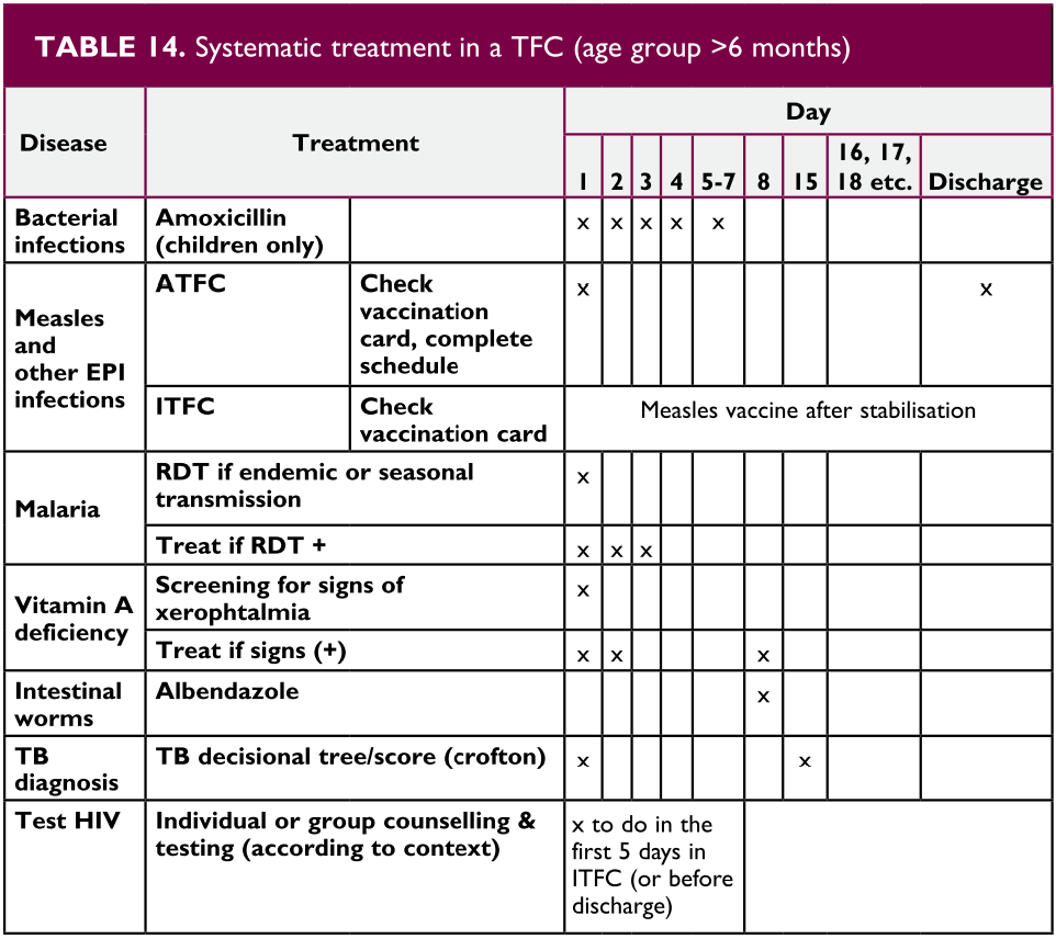
Table 14
6.4. Discharge Criteria
When using an ATFC manage children in their home with regular (ideally weekly) visits to the center and if possible a home visitor. The minimum stay in an ATFC program is 3 weeks. These children receive appropriate medical treatment for conditions that do not require hospitalization and RUTF. Infants 1 to 6 months of age can transfer to an ATFC when their medical complications have improved and they gain 10-15 g/kg/day for 5 consecutive days. Children 6 month to 10 years can be transferred to an ATFC or home when their edema has resolved; medical infections and other complications are resolving and no longer need oxygen, IV or IM treatment or close monitoring; the child’s appetite has normalized, and at least 2 weight measurements demonstrate an increasing trend. When an ATFC is not available ‘close to home” it is prudent to keep a child in the ITFC a little longer.
Children being managed in an ATFC or at home need admission or readmission to an ITFC if they develop signs of a severe medical complication with IMCI danger signs or a possible serious infection including HIV and TB. Nutritional reasons include development of edema, weight loss or poor weight gain (<5 g/ kg/day) after 2-3 weeks in the ATFC or home, failure of the appetite test.
Infants 1-6 months of age can be discharged from the ATFC when their W/H z score is > -2 on 2 consecutive visits and their weight is following their growth curve. Children 6 months to 10 years may be discharged from the ATFC when their MUAC is greater than 115 mm on 2 consecutive visits and their W/H z score is >/= -2 on 2 consecutive visits. Adolescents can be discharged when their W/H % is >80% on 2 consecutive visits.
7. Breastfeeding Programs in Disaster Situations
Objectives
- Recognize the importance of breastfeeding as well as its nutritional and logistic benefits in an emergency context.
Breast-feeding
Breast milk is the ideal form of nutrition for all infants during the first 6 months of life, making the consumption of other food resources by this age group unnecessary. The World Health Organization (WHO) recommends that breastfeeding be continued until the child is at least 24 months old, progressively supplemented with appropriate complementary foods after 6 months of age. In emergency settings where food supplies are limited, human milk remains an invaluable source of critical nutrients, particularly proteins. Therefore, it is important to provide adequate nutrition to lactating mothers.
Breast milk protects the infant against acute respiratory infections and diarrhea, both causing significant morbidity and mortality among infants and younger children. Overcrowded conditions, and limited access to clean and adequate water supplies, and stool disposal systems significantly increase the risk for these diseases in disaster situations. There is a common misconception that maternal stress or malnourishment leads to an inability to breastfeed. In fact, maternal hormone and neurotransmitter release during breastfeeding can help a mother to relax and attenuate stress and anguish caused by the disaster. The quality and quantity of breast milk has been shown to be adequate in all but the most severe degrees of maternal malnutrition. However, Vitamin D deficiency in the mother is associated with low Vitamin D in the breast milk, so supplementation is warranted in areas where mother’s have a low sun exposure due to cultural factors or where diet is low in Vitamin D.
In emergency settings where food supplies are limited, human milk remains an invaluable source of critical nutrients, particularly protein. Therefore, it is important to provide adequate nutrition to lactating mothers.
Introducing nonhuman milk or formula either to supplement or to complement breast milk, decreases maternal milk production to the point where it may compromise breastfeeding when a safe and sustainable supply of formula is no longer available.
Resources needed to safely feed a child non-human milk or formula, namely clean water, appropriate containers and methods for storage, and a safe and effective way to clean the containers or bottles, are always scarce in an emergency setting, as is the continued availability of formula or milk itself.
Consider the careful and judicious use of breast milk substitutes in special circumstances, provided replacement feeding is feasible, affordable, sustainable, and safe. Also consider breast milk substitutes in the case of orphans or children with mothers who were killed during the event. In these cases, a wet nurse may be an alternative to human milk substitutes. Studies show that it is usually safer and easier to give option B ART to the mother during breastfeeding than switch to formula feeds. Check with your national guidelines concerning breastfeeding by HIV positive mothers.
Maternal hormone and neurotransmitter release during breastfeeding can help a mother to relax and attenuate the stress and anguish caused by the disaster.
7.1. Feeding programs
In a disaster situation, feeding program options can range from a general feeding program to therapeutic and supplemental feeding programs. It is essential to ensure an equitable and appropriate distribution of available food supplies, with special emphasis on targeting the vulnerable groups. These programs must integrate local habits and preferences to the greatest possible extent.
It is important that feeding programs integrate local habits and preferences to the greatest possible extent.
General feeding programs
These types of programs distribute food to all people affected by the disaster. General feeding programs can be designed as complementary (providing some food items that are limited or not available) or supplementary (giving nutritional support to vulnerable groups) nutrition.
There are two commonly used forms of ration distribution: wet rations (which can be consumed without further preparation and are distributed at a feeding center) and dry rations (which require cooking and are consumed at the place of residence). There are advantages and disadvantages to each form of ration distribution.
Wet ration distribution ensures that the target individual consumes it, allows for the delivery of complementary health care services, and eliminates safety concerns that must be considered when carrying dry rations to the homes of children and women who can be victims of violence. In addition, when the food is prepared in a community site, participants do not need any fuel supply or cooking utensils. On the other hand, wet rations are often not feasible for large populations as they are labor-intensive and expensive.
Dry ration distribution is associated with lower cost and can reach larger numbers of individuals with fewer staff resources. Programs in Africa have introduced semisolid supplemental foods called ready to use therapeutic food (RUTF) that successfully improved early childhood nutritional status in malnourished populations. In addition, the family unit is maintained in their living area with the mother or caregiver spending less time away from the children. Feeding responsibility remains within the family, so that the habits and preferences of the affected population are more likely to be considered.
Recent experiences with cash transfers have had positive outcomes. These are often cheaper than providing food directly and do not disrupt local food markets. Cash transfer may also be more culturally acceptable and provide for a greater variety in the diet. Most of the time the money seems to be used to purchase food for the family.
8. Nutritional Status of Infants 0 to 6 Months of Age
Objectives
- Assess nutritional status and rule out feeding problems.
- Counsel the mother on effective breastfeeding.
- Know admission and discharge criteria for nutritional programs for children from 1-6 months of age.
Nutritional status and feeding problems
Assessing nutritional status and feeding problems during the first 6 months of life is a key aspect of health care. Detection of feeding problems and early diagnosis and treatment of infants with reduced weight gain or with weight loss may help prevent disease and death.
Causes of weight loss
A newborn can lose up to 10% of body weight during the first week of life due to edema reabsorption and fluid elimination. Weight loss is strongly conditioned by gestational age, birthweight, type and method of feeding and other factors associated with morbidity during the first days of life. Doctors Without Borders criteria for severe malnutrition indicating the need for admission to a Therapeutic Feeding Center or hospital include bilateral edema of the feet or for infants with a height less than 45 cm a confirmed weight loss of more than 10% of a prior documented weight. For infants with a height of 45-65 cm, criteria for admission is a W/H z score <-3. Severe acute malnutrition is considered complicated when there is also an identified medical complication. Medical complications include the presence of any danger sign and or a classification requiring hospitalization. Remember that infants under 2 months who present with signs of severe disease or local infection need inpatient care. In addition, there is consensus, that when infants younger than 6 months cannot feed because of breast feeding difficulties or inadequate formula feeding, they need admission regardless of their weight or presence of edema.
Detection of feeding problems and early diagnosis and treatment of infants with reduced weight gain or with weight loss may prevent disease and death.
Criteria for when to hospitalize or admit an infant to a therapeutic feeding center are found in Table 15.
| Criteria for Admission into an ITFC | |
|---|---|
| Nutritional status |
|
| Additional criteria | And/Or evidence of insufficient food intake |
| Criteria for Moving to an ATFC | |
|---|---|
| Nutritional status |
|
| Additional criteria | Plus ability to sustain appropriate feeding |
| Criteria for Discharge from the ATFC | |
|---|---|
| Nutritional status |
|
| After discharge | When discharged from the program, the infant is followed until the age of 6 months for growth monitoring, mother support and the provision of infant formula if needed. |
Weight loss during the first months of life has several causes, but it most frequently is related to feeding problems. Infants who have had repeated illnesses lose weight because of an altered appetite, foods, and caloric losses from vomiting or diarrhea. Infants who are not receiving adequate amounts of breast milk or appropriate alternatives for their age may have severe malnutrition or other nutritional disorders.
The newborn can lose up to 10% of body weight in the first week of life due to edema reabsorption and fluid elimination. The weight loss is strongly conditioned by gestational age, birth weight, type and method of feeding, and other factors associated with morbidity during the first days of life.
It is always important to counsel the mother on the appropriate breastfeeding technique and to encourage breastfeeding (Box 7).
BOX 7. Correct technique for breastfeeding (attachment and position)
- Check for lesions on the nipples
- Show the mother how to hold the infant
- With the body and head straight in one line
- With the body of the infant close to hers (belly contact)
- Holding the whole body of the child with her arm
- Show the mother how to help the infant get a good attachment while suckling
- Touch the infant’s lips with the nipple
- Wait until the infant opens his/her mouth wide
- Get the infant close to the breast quickly and make sure that the lower lip stays well under the nipple
- Check for signs of good attachment and suckling. If they are not good enough, try again
- Check that the mother is at ease
- Give support and reinforce the mother’s role
Weight loss during the first week of life should not exceed 10% of the birth weight. If the infant has lost more than 10 % of body weight, he/she will be considered to have a severe nutritional problem and must be immediately referred to a hospital to be evaluated by a specialist.
For infants whose weight loss does not exceed 10% of birth weight during the first week of life, weight for age will be evaluated by comparison to the weights of children the same age, using standard growth charts. Identify infants whose weight for age is under the lower percentile in the growth chart. These infants have a very low weight and need special care regarding their feeding.
It is also important to evaluate for good attachment and positioning during breastfeeding.
To verify good attachment, check for the following:
- The chin touches the breast (or very close)
- The mouth is wide open
- The lower lip is turned outward
- More areola is visible above than below the mouth
To verify the correct position, check if:
- The head and body of the infant are straight
- The infant’s head is directed to the mothers breast, with the nose in front of the nipple
- The infant’s body is close to the mother’s body (belly-to-belly)
- The mother is holding firmly the entire body of the infant, not just the shoulders and neck.
9. Summary
Adequate nutrition is vital to everyone’s health and well-being. Even in the best of times there are multiple challenges to proper nutrition. These challenges are greatly increased in the aftermath of a natural or man-made disaster. An understanding of the local community and reliable information on local resources are critical in the development of a recovery strategy. It is important to remember that malnutrition increases morbidity and mortality of the affected population, particularly among the most vulnerable groups, such as children. Assessing the nutritional status of the population (through anthropometric measurements), identifying macro- and micronutrient deficiencies, and implementing preventive and therapeutic strategies increase dramatically the likelihood of successful recovery among populations affected by a disaster. Under these circumstances, the IMCI primary health care strategy provides a reasonable approach to enhance the achievement of this goal.
10. Suggested Reading
References
- Academy for Educational Development. Recommended Feeding and Dietary Practices to Improve Infant and Maternal Nutrition. Washington, DC: Academy for Educational Development; 1999.
- Basch PF. Textbook of International Health. New York: Oxford University Press; 1999.
- BASICS II. Nutrition Essentials: A Guide for Health Managers. Washington, DC: BASICS; 2004.
- Boelaert M, Davis A, et al. Nutrition Guidelines. Paris: Médecins sans Frontières; 1995.
- Bread for the World Institute. Countries in Crisis: Sixth Annual Report on the State of World Hunger. Silver Spring, Md; 1995.
- Caballero B. A nutrition paradox—underweight and obesity in developing countries. N Engl J Med 2005;352:1514-1516.
- Canahuati J, et al. Infant and Young Child Feeding in Emergencies; Nutrition Module for the InterAction Health Training Curriculum, Academy for Educational Development; 1997.
- Cogill B. Nutrition in Emergency Situations; Impact Project. Paper presented at the Department of the Army Brooke Army Medical Center shortcourse on “Nutrition Support for Combat Casualties and Humanitarian Missions.” San Antonio, Texas; 1997.
- CORE. Positive Deviance/Hearth: A Resource Guide for Sustainably Rehabilitating Malnourished Children, 2003.
- D’Souza R. Vitamin A for the treatment of children with measles: a systematic review. J Tropical Pediatr 2002;48:323-327.
- Forbes GB, ed. Pediatric Nutrition Handbook. Elk Grove Village, Ill: American Academy of Pediatrics; 1985.
- Grobler-Tanner C, Collins S. Community therapeutic care (CTC): a new approach to managing acute malnutrition in emergencies and beyond. Food and Nutrition Technical Assistance. Technical Note Nº 8. June 2004.
- Hanson L. Vitamin A and intestinal function. In: Bhutta ZA, ed. Contemporary Issues in Childhood Diarrhea and Malnutrition. New York: Oxford University Press; 2000.
- Huffman SL, Baker J, et al. The Case for Promoting Multiple Vitamin/Mineral Supplements for Women of Reproductive Age in Developing Countries. Washington, DC: Academy for Educational Development; 1998.
- Humphrey J, West K Jr, et al. Vitamin A deficiency and attributable mortality among under-5-year-olds. Bull WHO 1992;70:225-232.
- Institute of Medicine. Prevention of Micronutrient Deficiencies: Tools for Policymakers and Public Health Workers. Washington, DC: National Academy Press; 1998.
- Institute of Medicine. High-Energy, Nutrient-Dense Emergency Relief Food Products. Washington, DC: National Academy Press; 2002.
- Institute of Medicine. Vitamin C Fortification of Food Aid Commodities. Washington, DC: National Academy Press; 1997.
- International Life Sciences Institute. Preventing Micronutrient Malnutrition: A Guide to Food-Based Approaches—A Manual for Policy Makers and Programme Planners. International Life Sciences Institute; 1997.
- Kelly M. Infant feeding in emergencies. Disasters1993;17:110-119.
- Khara T, Collins S. Emergency Nutrition Network (ENN) Special Supplement: Community-based Therapeutic Care (CTC). ENN Special Supplement Series, Nº 2. 2204.
- Mc Laren DS. A Colour Atlas and Text of Diet-Related Disorders. St. Louis, Mo: Mosby Year Book; 1992.
- Mears C, Chowdhury S. Health Care for Refugees and Displaced People. Oxfam Practical Health Guide No. 9; 1994.
- Mejia L, Chew F. Hematological effect of supplementing anemic children with vitamin A alone and in combination with iron. Am J Clin Nutr 1988;48:595-600
- Merck Publications. The Merck Manual Online. Section 11, Hematology and Oncology. Chapter 130, Anemias. 2006.
- Merck Publications. The Merck Manual Online. Section 1, Nutritional Disorders. Chapter 4, Vitamin Deficiency, Dependency, and Toxicity. 2006. http://www.merck.com/mmpe/sec01/ch004/ch004a.html.
- Merson M, Black RE, et al. International Public Health: Diseases, Programs, Systems, and Policies. Gaithersburg, Md: Aspen Publishers; 2001.
- Refugee Health Unit, Somali Ministry of Health. Guidelines for Health Care in Refugee Camps. Oxford; 1983.
- Saps M, Heyman MB. Nutrition. In: Hyman PE, ed. Pediatric GI Problems. Philadelphia, Pa: Current Medicine; 1997.
- Semba R, Bloem M. Nutrition and Health in Developing Countries. Totowa, NJ: Humana Press; 2001.
- Shils ME, Shike M, Ross AC, et al. Modern Nutrition in Health and Disease. 10th edition. London: Lippincott Williams & Wilkins; 2006.
- Solomon SM, Kirby DF. The refeeding syndrome: a review. J Parenter Enteral Nutr 1990;14: 90-97.
- Sommer A, West K Jr. Vitamin A Deficiency: Health, Survival, and Vision. New York: Oxford University Press; 1996.
- Sphere Project. Humanitarian Charter and Minimum Standards in Disaster Response. Geneva: Oxfam Publishing; 2004.
- Suharno D, West C, et al. Supplementation with vitamin A and iron for nutritional anaemia in pregnant women in West Java, Indonesia. Lancet 1993;342:1325-1328.
- Suskind RM, Lewinter-Suskind L. Textbook of Pediatric Nutrition. 2nd ed. New York, 1993.
- Thurnham DI. Micronutrients and immune function: some recent developments. J Clin Pathol 1997;50:887-891.
- United Nations Children’s Fund. Preventing Iron Deficiency in Women and Children: Background and Consensus on Key Technical Issues and Resources for Advocacy, Planning and Implementing National Programmes. UNICEF/UNU/WHO/MI Technical Workshop. New York: UNICEF; 1998.
- USAID. Nutritional Management of Malnourished Children.
- Walker WA, Watkins JB, et al. Nutrition in Pediatrics: Basic Science and Clinical Applications. Hamilton, Ontario: BC Decker; 2003.
- Waterlow JC. Protein Energy Malnutrition. London; 1992.
- West K Jr, Darnton-Hill I. Vitamin A deficiency. In: Semba R, Bloem M, eds. Nutrition and Health in Developing Countries. Totowa, NJ: Humana Press; 2001.
- West K Jr, Rice A, et al. Tables on the Global Burden of Vitamin A Deficiency Among Preschool Children and Women of Reproductive Age.
- WHO/UNICEF. (2004). WHO/UNICEF Joint Statement: Clinical Management of Acute Diarrhea.
- WHO/UNICEF. Oral Rehydration Salts (ORS) A New Reduced Osmolarity Formulation, 2002.
- World Health Organization. Management of Severe Malnutrition: A Manual for Physicians and Other Senior Health Workers. Geneva: WHO; 1999.
- World Health Organization. Management of the Child with a Serious Infection or Severe Malnutrition: Integrated Management of Childhood Illness. Geneva: WHO; 2000.
11. Case Resolution
Case 1.
In order to collect information on the nutritional status of the affected population prior to the disaster, it would be useful to get in contact with the local public health authorities and health professionals. It is also necessary to collect information regarding the most vulnerable groups (e.g., people included in pre-existing food programs). Lastly, it is necessary to have information regarding the available food resources in order to develop the most appropriate and sustainable distribution programs for the aftermath of the disaster.
Case 2.
Since this is a preterm infant, it is important to ensure adequate iron intake earlier than in term infants. At 4 months of age the infant should not be receiving any supplementary foods in addition to breastfeeding. The infant should therefore be supplemented with iron at a dose of 2 mg/kg/day.
Case 3.
You must first look for signs indicating malnutrition and anemia. There is no serious wasting or edema in the lower extremities. The palms are slightly pale. The anthropometric measurements show a 40% weight/height deficit when compared with standard weight/height growth charts: very low weight for age. He is therefore classified as having severe malnutrition and must be referred to a hospital for proper treatment. In these children, the therapeutic refeeding program must be careful to avoid the refeeding syndrome and the associated increased morbidity and mortality.
12. Book Review
SECTION I - NUTRITIONAL STATUS ASSESSMENT
- How would you assess the nutritional status of a population affected by a disaster?
- Which are the most vulnerable populations in an emergency setting?
- Which anthropometric measurements are used to assess nutritional status in children?
SECTION II - CLINICAL PRESENTATION OF MALNUTRITION
- What are the clinical features of severe acute malnutrition?
- What are the physiological features in severe malnutrition that increase the risk for the refeeding syndrome?
- What are the clinical and physiological manifestations of the refeeding syndrome?
SECTION III - MICRONUTRIENT DEFICIENCIES
- What are the epidemiological and clinical implications of iron, vitamin A, and zinc deficiencies?
- Which interventions allow the prevention or correction of micronutrient deficiencies in a population affected by a disaster?
SECTION IV - IMCI STRAGEGY FOR NUTRITIONAL STATUS ASSESSMENT
- What elements are used to classify malnutrition and anemia according to the IMCI guidelines?
- What factors determine the appropriate management for each category?
SECTION V - FEEDING PROGRAMS IN DISASTER SITUATIONS
- What are the logistic and nutritional benefits of breast-feeding?
- What are the goals and characteristics of the different feeding programs that can be used in these situations?
- What are the phases of a therapeutic feeding program?
SECTION VI - NUTRITIONAL STATUS OF INFANTS 0 TO 6 MONTHS OF AGE
- What is the expected change in body weight for the newborn during the first days of life?
- Which elements are included in the assessment of the nutritional status in infants less than 2 months of age?
- What are the characteristics of a good attachment and position during suckling?
13. Appendix
Treatment of Hypophosphatemia
| Degree of Hypophosphatemia | IV Phosphate Replacement Dosage Administer over 6-12 hours |
|---|---|
| 2.3-2.7 mg/dL, asymptomatic | 0.08-0.16 mmol/kg |
| 1.5-2.2 mg/dL, asymptomatic | 0.16-0.32 mmol/kg |
| <1.5 mg/dL, symptomatic | 0.32-0.64 mmol/kg |
Note: Ensure adequate renal function: in patients with renal insufficiency give 50% initial dose Adapated from Refeeding Syndrome. Pediatr Clin N Am 56 (2009)1201-1210.
Treatment of Hypokalemia
| Degree of Hypokalemia | IV Potassium Replacement Dosage Administer over at least 1 hour |
|---|---|
| 2.5-3.4 mEq/L | 0.3-0.4 mEq/kg per dose over at least 1 hour |
| <2.5 mEq/L | 0.4-0.5 mEq/kg per dose over at least 1 hour |
Note: Ensure continuous cardiorespiratory monitoring Adapated from Refeeding Syndrome. Pediatr Clin N Am 56 (2009)1201-1210.
Treatment of Hypomagnesemia
| Degree of Hypomagnesemia | IV Magnesium Replacement Dosage Administer over 4 hours |
|---|---|
| 1-1.5 mg/dL | 0.2-0.3 mEq/kg per dose |
| <1 mg/dL | 0.3-0.4 mEq/kg per dose |
Note: Ensure adequate renal function: in patients with renal insufficiency give 50% initial dose One gram magnesium sulfate = 8.1 mEq magnesium Adapated from Refeeding Syndrome. Pediatr Clin N Am 56 (2009)1201-1210.
13.1. Appetite Test
This is one of the criteria for deciding if a patient should receive outpatient or inpatient hospital care. Poor appetite means a child is suffering from a serious infection and/or metabolic disorder. These patients’ lives are at risk, and must be referred to the inpatient therapeutic feeding center or hospital.
The child should be able to take the minimum amount required to maintain body weight. S/he should not be sent home if there is any risk of continued deterioration, on account of not taking enough RUTF.
How to carry out the appetite test
- The appetite test should be carried out at each visit for out-patients (particularly those who do not gain weight)
- It should be carried out carefully, in a quiet place.
- Explain the purpose of the test to the caregiver.
- S/he should wash his/her hands, as well as the child’s hands and face, sit comfortably and take the child on his/her lap.
- The RUTF should be offered either directly from the sachet, or by putting a small amount on the caregiver’s finger (the first approach is preferable in order to reduce the risk of contamination). The caregiver should offer the child the RUTF gently, without forcing him/her, and encourage the child regularly. If the child refuses it, the caregiver should continue to quietly encourage the child and take the time needed.
- For the children < 1 year and where the BP100 is used, make porridge by adding some water to the biscuit.
- The test is generally short, but may last up to 1 hour. The child must never be forced to take the RUTF.
- Drinking water must be given at the same time ( the RUTF paste/biscuit is thick and inedible without sufficient water)
After 1 hour, the child should have eaten at least:
| Peanut paste (sachet of 92g) | |
|---|---|
| Minimum quantity | ⅛ to ¼ of the sachet |
| BP 100 (Bars) | |
|---|---|
| Not applicable | - |
| Peanut paste (sachet of 92g) | |
|---|---|
| Minimum quantity | ¼ to ⅓ of the sachet |
| BP 100 (Bars) | |
|---|---|
| Minimum quantity | ¼ to ½ |
| Peanut paste (sachet of 92g) | |
|---|---|
| Minimum quantity | ¼ to ⅓ of the sachet |
| BP 100 (Bars) | |
|---|---|
| Minimum quantity | ½ to ¾ |
| Peanut paste (sachet of 92g) | |
|---|---|
| Minimum quantity | ⅓ to ½ of the sachet |
| BP 100 (Bars) | |
|---|---|
| Minimum quantity | ½ to ¾ |
| Peanut paste (sachet of 92g) | |
|---|---|
| Minimum quantity | ½ to ¾ of the sachet |
| BP 100 (Bars) | |
|---|---|
| Minimum quantity | ¾ to 1 |
| Peanut paste (sachet of 92g) | |
|---|---|
| Minimum quantity | ¾ to 1 sachet |
| BP 100 (Bars) | |
|---|---|
| Minimum quantity | 1 to 1½ |
| Peanut paste (sachet of 92g) | |
|---|---|
| Minimum quantity | >1 sachet |
| BP 100 (Bars) | |
|---|---|
| Minimum quantity | >1½ |
The appetite test provides an ideal opportunity to observe mother-child interaction, detect any psychosocial problems and carry out health promotion activities.
Result of the appetite test
The result of the appetite test is considered to be “good or average” if the child takes approximately the volume (or more) indicated in the table above.
The appetite test failed if the child takes less than the volume of RUTF indicated in the table above. The child must then be referred to the hospital or inpatient therapeutic feeding center.