Management of Prevalent Infections in Children Following a Disaster
| Site: | Pediatric Pandemic Network Learn |
| Course: | Management of Prevalent Infections in Children Following a Disaster |
| Book: | Management of Prevalent Infections in Children Following a Disaster |
| Printed by: | Guest user |
| Date: | Saturday, September 12, 2026, 5:25 PM |
Table of contents
- 1. Overview
- 2. Introduction
- 3. Integrated Management of Childhood Illness (IMCI)
- 4. Influenza Infections
- 4.1. Epidemiology
- 4.2. Influenza Pathogenesis and Symptoms
- 4.3. Complications of Influenza
- 4.4. Diagnostic Tests
- 4.5. Treatment of Influenza
- 4.6. Chemoprophylaxis
- 4.7. Prevention of Influenza
- 4.8. Surveillance and Surge Planning
- 4.9. Special Issues in Developing Countries
- 4.10. Lessons Learned from 2009-2010 H1N1 Pandemic
- 5. Measles
- 6. Acute Respiratory Infections
- 7. Febrile Illnesses
- 8. Other Cases That Require Attention at the Scene of the Disaster
- 9. Vaccination in Disaster Situations
- 10. Infections in Infants 0 to 2 Months of Age
- 11. Summary
- 12. Suggested Reading
- 13. Case Resolution
- 14. Book Review
- 15. Appendix
1. Overview
Welcome to this multimedia open access edition of this chapter book titled Management of Prevalent Infections in Children Following a Disaster Book.
This content is available as a digital book in two formats:
1) Website, which you are currently reading. This format offers a mobile friendly multimedia experience with sharable links to chapters. There are three ways to move through the book:
a) Use the table of contents chapter navigation to the left to navigate to different chapters in the book. Scroll through the links to select the chapter that interests you.
b) Use the yellow arrows that appear when scrolling to select previous and next chapters.

c) Use the previous chapter and next chapter buttons at the bottom of very page to navigate through the book.

2) Printable, which you can save as a PDF and open without an Internet connection.
1.1. Acknowledgements
Created by the Center for Global Health, Colorado School of Public Health
This content is owned by the Center for Global Health, Colorado School of Public Health, and has been jointly created by the Center for Global Health, Colorado School of Public Health and the Maternal and Child Health Department of Maimonides University; both of which are WHO Collaborating Centers in Maternal and Child Health. The course materials were developed with input from the American Academy of Pediatrics (AAP), the Pan American Health Organization (PAHO), and the Association for Health Research & Development (ACINDES).
With Support from the Pediatric Pandemic Network

The Pediatric Pandemic Network is supported in part by the Health Resources and Services Administration (HRSA) of the U.S. Department of Health and Human Services (HHS) as part of cooperative agreements U1IMC43532 and U1IMC45814 with 0 percent financed with nongovernmental sources. The content presented here is that of the authors and does not necessarily represent the official views of, nor an endorsement by HRSA, HHS, or the U.S. Government. For more information, visit HRSA.gov.

Attribution-NonCommercial-NoDerivatives CC BY-NC-ND
You are free to download and share this work for noncommercial purposes, as long as you credit the Center for Global Health and the Pediatric Pandemic Network for the original creation.
2. Introduction
Book Authors
Stephen Berman, MD, FAAP | Ribka Amsalu, MD, MSc | Nee-Kofi Mould-Millman, MD | Lisa L. Abuogi, MD, MSc | Chris Nyquist, MD, MSPH | Col. Julia A. Lynch, MD, FAAP | Ángela Gentile, MD | Daniel Martinez Garcia, MD, MPH
Morbidity and mortality resulting from an acute humanitarian emergency in developing countries are related to the excessive childhood mortality that existed prior to the disaster. The main causes of mortality are — neonatal causes, pneumonia, diarrhea — followed by malaria, measles, injury, and meningitis. Malnutrition is an underlying condition that increases the risk of dying from all of the above causes.
During acute humanitarian emergencies, mortality related to those common childhood infections increases due to crowded living conditions; displacement to areas with higher disease prevalence; and compromised personal hygiene resulting from inadequate water supplies, contaminated water, and poor sanitation. The preexisting nutritional status (particularly micronutrient and vitamin A deficiencies) and immunization rates of children, as well as the pre-existing primary care infrastructure and the degree of damage caused by the disaster, also affect childhood morbidity and mortality after a disaster. Figure 1 summarizes the causes of death in two refugee camps and illustrates the significance of various illnesses following a disaster.
Figure 1. Mortality in Two Refugee Camps
Figure 1. Mortality in two refugee camps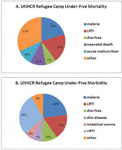
|
Figure 1. Cause-specific morbidity and mortality in children younger than five years of age for refugee camps in the UNHCR HIS database, January 2006 to February 2010. A. Mortality for all recorded causes. Cases of watery and bloody diarrhea were combined. B. Out-patient visits for major causes of morbidity within refugee camps. Suspected and confirmed cases of malaria were combined, as were cases of watery and bloody diarrhea. Only causes accounting for 5% or more of mortality or morbidity are shown, with the remaining causes listed as “Other”. |
These stark statistics emphasize the importance of using strategies based on the Integrated Management of Childhood Illness (IMCI) Program, developed to prevent and treat infections during an acute humanitarian emergency.
3. Integrated Management of Childhood Illness (IMCI)
OBJECTIVES
- Describe the rationale for the WHO evidence-based syndromic approach to case management as described in the IMCI.
- List the clinical illnesses included in the IMCI program and their relevance in situations associated with disasters.
- Assess and classify the condition of a child to determine its severity and establish the relationship between this classification and the subsequent management.
- List the danger signs that should be routinely checked in all children.
What is Integrated Management of Childhood Illness (IMCI)?
The strategy for the IMCI was designed by WHO to enhance children’s health and reduce mortality and morbidity caused by the most prevalent childhood diseases throughout the world, but especially in countries having the highest under 5 mortality rates. The IMCI strategy addresses most, but not all, of the major reasons why a sick child needs medical attention. A child with a chronic condition or a less common illness may require additional special care and IMCI guidelines do not describe the management of trauma or other acute emergencies due to accidents or injuries.
Case 1:
A 15-month-old boy presents at the emergency department with a fever. He had been healthy until 3 days ago, when he developed symptoms of upper respiratory airway infection. His mother reports giving ibuprofen to her son the day before, because of the fever. The child continues to be febrile with reduced food and fluid intake, urine output, and activity level. There is no history of vomiting, diarrhea, cough, or rash. He is not receiving any medication. You note fatigue and irritability when the child is stimulated during the physical examination. Respiratory rate is 50 breaths/min, pulse rate 162 beats/min, blood pressure 92/70 mm Hg, and axillary temperature 38.9ºC. He has dry lips but wet oral mucosa without lesions. His neck is flexible. Lung and heart examination are unremarkable with no significant findings. A few isolated petechiae are noted over the abdomen and lower limbs. Peripheral pulse is normal and capillary refill time is 3 seconds.
- What is your global clinical impression for this boy?
- What is the most probable diagnosis?
- What treatment strategies you should adopt initially?
This IMCI strategy seeks to integrate the acute care for children under 5 years of age across the continuum of clinical services; from community health worker encounters through first level referral health care facilities (camps, medical offices, health care centers and hospital primary care departments) and then in hospitals. The clinical decision making approach involves using a limited number of symptoms and signs to classify the severity of illness, which determines the child’s treatment/management. See Figure 2.
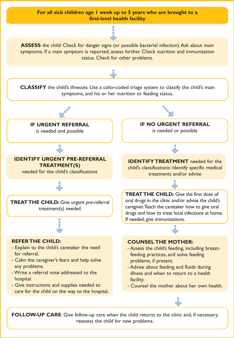
Figure 2. Summary of the process of integrated care of children
IMCI management also includes guidelines for follow-up, counseling the parents, and instructions regarding when to return when additional care is needed. The IMCI approach focuses training and materials on the need to improve parental skills and practices related to care seeking behaviors, the value of preventive services such as immunization and nutrition, and home care of children. The basic principles of IMCI are presented in Box 1.
BOX 1: Principles of the Integrated Clinical Case Management
IMCI clinical guidelines are based on the following principles:
- Examining all sick children aged up to five years of age for general danger signs and all young infants for signs of very severe disease. These signs indicate severe illness and the need for immediate referral or admission to hospital.
- The children and infants are then assessed for main symptoms:
- In older children the main symptoms include:
- Cough or difficulty breathing,
- Diarrhoea
- Fever, and
- Ear infection
- In young infants, the main symptoms include:
- Local bacterial infection,
- Diarrhoea, and
- Jaundice,
- In older children the main symptoms include:
- Then in addition, all sick children are routinely checked for:
- Nutritional and immunization status,
- HIV status in high HIV settings, and
- Other potential problems.
- Only a limited number of clinical signs are used, selected on the basis of their sensitivity and specificity to detect disease through classification.
A combination of individual signs leads to a child’s classification within one or more symptom groups rather than a diagnosis. The classification of illness is based on a colour coded triage system:
- “PINK” indicates urgent hospital referral or admission,
- “YELLOW” indicates initiation of specific outpatient treatment.
- “GREEN” indicates supportive home care.
- IMCI management procedures use a limited number of essential drugs and encourage active participation of caregivers in the treatment of their children.
- An essential component of IMCI is the counselling of caregivers regarding home care:
- Appropriate feeding and fluids,
- When to return to the clinic immediately, and
- When to return for follow-up
3.1. IMCI Guidelines
The IMCI charts can be found in the appendix of this module.
The IMCI strategy is divided into 2 components based on the child’s age; management of the sick child aged 2 months up to 5 years and management of the sick young infant aged up to 2 months.
The sick child management includes respiratory disease, diarrhea, febrile illness (malaria), measles, ear infections, malnutrition, anemia, HIV, and immunization status. See Figure 3.
Figure 3. Management of the Sick Child Aged 2 Months Up to 5 Years
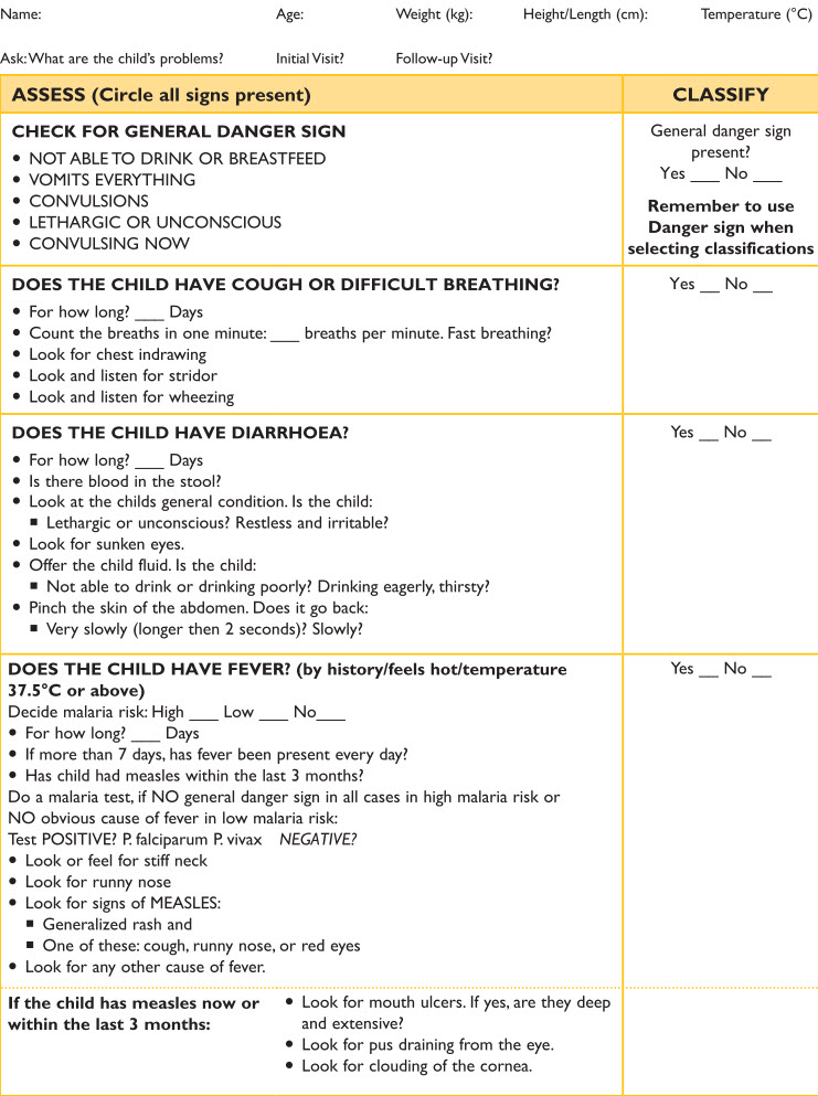
Figure 3. Management of the Sick Child Aged 2 Months Up to 5 Years
Figure 3. Management of the Sick Child Aged 2 Months Up to 5 Years, Continued
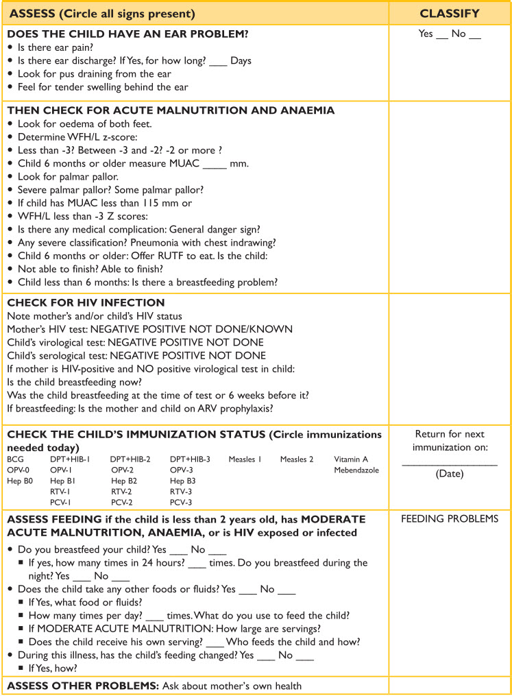
Figure 3. Management of the Sick Child Aged 2 Months Up to 5 Years, Continued
The sick young infant management includes severe disease (respiratory and sepsis), local bacterial infection, jaundice, diarrhea, HIV infection, weight gain, breast feeding and other feeding problems, immunization status and mother’s health. See Figure 4.
Figure 4. Management of the Sick Young Infant Aged Up to 2 Months
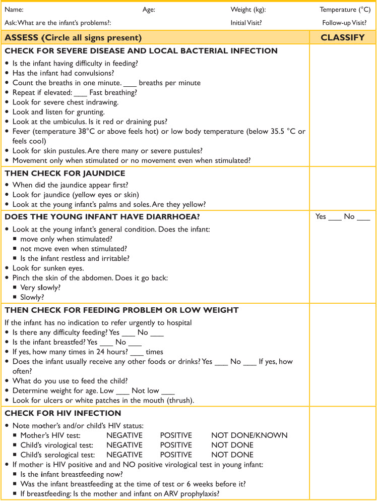
Figure 4. Management of the Sick Young Infant Aged Up to 2 Months
Figure 4. Management of the Sick Young Infant Aged Up to 2 Months, continued
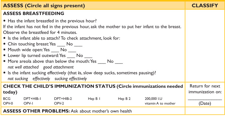
Figure 4. Management of the Sick Young Infant Aged Up to 2 Months, continued
IMCI related research has identified from the traditional clinical history and physical examination a limited number of clinical symptoms and signs, based on evidence of their sensitivity and specificity to detect diseases and predict mortality.
These were selected also considering the available resources in first-level health care facilities. Obtaining the necessary information uses a ask, look and listen training approach. The presence of these more limited symptoms and signs leads to a child’s classification rather than a diagnosis. This classification indicates the severity of the condition, which calls for specific actions based on whether the child (a) needs to be urgently referred to a higher level of care, (b) requires specific treatments, or (c) can be safely managed at home. The classification is color coded: pink requires hospital referral or admission; yellow indicates the need to initiate treatment at home; and green indicates home supportive care management.
Assessment of sick children 2 months to 5 years
The assessment procedure for this age group includes a number of important steps that must be taken by the health care provider: (1) Take a history and talk with the caregiver about the child’s problem; (2) check for general danger signs; (3) assess major symptoms; (4) evaluate nutritional status; (5) assess the child’s feeding; (6) check immunization status; and (7) look for other problems.
Danger signs that should be routinely checked in all children
In the sick child management there are 5 general danger signs: 1. not able to drink or breast feed, 2. vomits everything, 3. convulsions, 4. lethargic or unconscious, and 5. now convulsing. These general danger signs indicate the need for immediate referral to a hospital because they predict serious infections and conditions such as bacterial meningitis, cerebral malaria, and septic shock, which untreated often lead to death.
Seizures are associated with meningitis, cerebral malaria, or other life threatening conditions. However, not all seizures may be life threatening, as some seizures may result from fever. Febrile seizures do little harm beyond frightening the parents.
An unconscious child is likely to be seriously ill. A lethargic child who is awake but does not take any notice of his/her surroundings or does not respond normally to sounds or movement may also be very sick. These signs can be associated with many conditions, including dehydration, meningitis.
Inability to drink or breastfeed. An infant may be unable to drink if he/she is too weak or cannot swallow. Observe the child while the mother breastfeeds or gives him/her something to drink.
Persistent vomiting. Vomiting itself may be a sign of serious illness. This symptom may also prevent the child from taking medications or fluids for rehydration.
A child with one or more of these signs must be considered seriously ill and will require hospital referral. To start treatment for severe illnesses without delay, quickly assess the child for the most important causes of serious illness and death, including acute respiratory infection (ARI), diarrhea and dehydration, sepsis, malaria, and measles.
3.2. Cough and Difficult Breathing
When a child presents with cough, the parent is asked about the presence of fast breathing. Fast breathing results when lung compliance is reduced by infiltrate or consolidation associated with pneumonia or bronchiolitis. Research has established respiratory rate (breaths per minute) thresholds for fast breathing: 60 in an infant up to 2 months, 50 in child 2 months to 12 months, and 40 in a child 12 months to 5 years. When the child is calm, respirations should be counted for 1 minute. One minute timers are available to facilitate the accurate counting of breaths/minute. Subcostal chest indrawing (retractions) is associated with decreased oxygen saturation that affects the central nervous system respiratory drive center, which controls the force of the diaphragm. In addition to checking for fast breathing and chest indrawing, it is important to listen for stridor and wheezing. The child is classified having severe pneumonia or another very severe disease (PINK) if there is any general danger sign or stridor in a calm child. This child needs to be referred urgently to the hospital and receive a first dose of an antibiotic. If the child has fast breathing and or chest indrawing , the child is classified as having pneumonia (YELLOW) and is given oral amoxicillin (or an appropriate alternative) for 5 days. Wheezing children are given a trial with a rapid acting bronchodilator for up to 3 times. Children without any signs of pneumonia or danger signs are classified as having an upper respiratory infection or cold (GREEN) and receive home care with a safe cough remedy. If their wheezing responded to a therapeutic rapid acting bronchodilator trial they are given a rapid acting bronchodilator for 5 days. (See IMCI chart for cough or difficult breathing in the appendix.)
Diarrhea
When a child presents with diarrhea, the classification of severity is based on whether there is blood in the stool, how long the child has had diarrhea, and the presence of the following clinical signs: unconscious, lethargy, restless, irritable, sunken eyes, ability and way of drinking, skin turgor. These signs are used to classify the severity of dehydration. IMCI management of diarrhea is reviewed in module 6.
Fever and Measles
When a child presents with fever by history or a temperature higher than 37.5 C., the severity classification is based on the duration of the fever, whether the child had measles within the past 3 months, and the following signs: a stiff neck, runny nose, any bacterial cause of the fever, signs of measles. The classification takes into account whether the child lives in an area with malaria and if the child now has measles or had within the last 3 months. The management of malaria and measles is reviewed later in this module. (See IMCI chart for fever in the appendix)
Ear infection
When a child presents with ear pain or ear discharge the classification considers if there is tender swelling behind the ear suggesting mastoiditis. A child with mastoiditis is classified as PINK and referred urgently to a hospital with the first dose of an appropriate antibiotic. Acute ear pain or ear discharge is considered yellow and the ear infection is treated with an antibiotic for 5 days. When the ear drainage has lasted more than 14 days the ear infection has become chronic and is managed by ear wicking and topical quinolone eardrops for 14 days. (The IMCI chart on ear infection is found in the appendix)
Malnutrition and Anemia
The child is assessed for acute malnutrition based on finding edema of both feet, the weight for length percentile ( Z score) using WHO growth standards and the mid upper arm circumference for children older than 6 months. Anemia is recognized by palmar pallor. The severity classification is made on the basis of these findings plus the presence of any medical problem/complication identified earlier in the IMCI decision making process. The IMCI management of malnutrition and anemia is reviewed in module 8.
HIV infection
In areas of the world with a high prevalence of HIV infection, IMCI checks for a possible HIV infection noting the mother and child’s HIV status ( virology test and serology test). The IMCI approach to HIV infection is reviewed later in this module.
Immunization Status, Feeding and Mother’s Health
IMCI then checks the child’s immunization status and assesses feeding if the child is less than 2 years old.
Other Problems
Asking about other parental concerns and problems is helpful. Mother’s health is always valuable as this has a direct impact on the child’s health.
3.3. Management of the Sick Young Infant up to 2 Months (see chart in the appendix)
Severe Disease and Local Bacterial Infection
Young children having any of the following danger signs have very severe disease and should be urgently referred to the hospital with a first antibiotic dose and treatment to prevent low blood sugar: 1. not feeding, 2. convulsions, 3. fast breathing ( more than 60 breaths per minute) 4. severe chest indrawing, 5. fever or low temperature, 6. lack of movement. A young infant with signs of umbilical infection (redness and or purulent discharge) is classified as yellow having a local bacterial infection and treated with an appropriate antibiotic).
Jaundice
An infant is assessed for jaundice by asking when the infant first looked yellow and noting what areas of the body appear yellow especially the soles and palms. The infant is classified as severe if the jaundice was noticeable before 24 hours of age or involves the soles and palms at any age. Jaundice is reviewed in module 7.
Diarrhea
When the infant presents with diarrhea the infant’s general condition and movements are noted as well as the presence of sunken eyes and skin turgor in order to classify the severity of dehydration. This is reviewed in module 6.
Poor weight gain, low weight, feeding problem
The infant’s classification is based on a feeding history related to breast feeds, additional foods or drinks, weight for age, and the presence of ulcers or white patches in the mouth suggesting thrush. Poor weight gain and breast feeding problems are reviewed in module 8.
HIV infection
In areas of the world with a high prevalence of HIV infection, IMCI checks for a possible HIV infection noting the mother and child’s HIV status ( virology test and serology test). The IMCI approach to HIV infection is reviewed later in this module.
Assess Breastfeeding
Observe the mother breastfeeding if the infant has not fed in the previous hour and check attachment and suck. Breastfeeding is reviewed in module 8.
Immunization Status and Mother’s Health
It is always necessary to ask about immunization status and assess for other problems such as mother’s health especially possible mastitis or severe postpartum depression.
Summary
MCI is family and community centered. This approach is essential for childhood health, because it promotes healthy habits in the family, adequate care of children (feeding, clothing, stimulation, etc.), disease prevention, and prompt seeking of medical care when alarming signs and symptoms are noted.
The IMCI strategy also helps healthcare professionals take advantage of opportunities for prevention, promote childhood development, and encourage the rational use of drugs and medications.
Figure 5. IMCI strategy for case management in the outpatient health care facility, first-level referral service, and at home for the sick child from age 2 months to 5 years
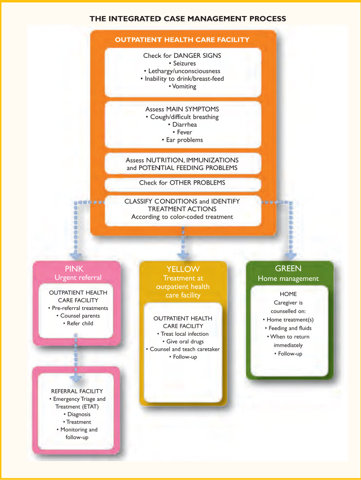
Figure 5. IMCI strategy for case management in the outpatient health care facility, first-level referral service, and at home
4. Influenza Infections
OBJECTIVES
- Understand the influenza virus pathogenesis and epidemiology.
- Describe clinical symptoms of influenza and diagnostic and treatment options.
- Evaluate treatment and prevention methodologies for influenza and incorporate them into preparedness plans.
What is Influenza?
Influenza is a segmented, single-stranded enveloped RNA virus classified into influenza A, B and C based on antigenic differences. Influenza A is a potentially severe illness, causes epidemics and pandemics, is rapidly changing, and infects birds, swine, horses, seals, and humans. Influenza B is more uniform, causes epidemics and only infects humans. Influenza C is of minimal public health impact and infects humans and swine. Further subtyping of influenza A virus is based on the neuraminidase and hemagglutinin proteins on the viral surface. There are 16 different hemagglutinins and 9 different neuraminidase subtypes. Hemagglutinin proteins allow the virus to stick to cells by binding to a specific receptor. The neuraminidase protein helps newly formed viral particles get released from the cell surface so that they have the potential to infect other cells. Only H1N1, H2N2, H3N2 subtypes are associated with widespread epidemics in human. Since 1997, rare but severe infections in humans with influenza A subtype H5N1 viruses have been identified in Asia, Africa, Europe, and the Middle East where these viruses are present in domestic or wild birds.
Repeated seasonal influenza epidemics persist because the type A and type B viruses undergo constant and rapid change due to antigenic drift. Antigenic drift refers to a gradual change in the virus that occurs through a slow series of amino acids changes in the hemagglutinin or neuraminidase surface antigens. Occurring only after a particular viral strain has become established in humans, antigenic drift represents an adaptation to the development of host antibodies. Newly developed antigenic strains of influenza then prevail for a period of 2 to 5 years, only to be replaced by the next emerging strain. This new strain can then trigger a new epidemic, since it is now unfamiliar to the antibody repertoire of the population. The development of yet another set of host antibodies eventually protects the population–at the same time it puts pressure on the virus to drift yet again. Ongoing change caused by antigenic drift requires ongoing reformulation of influenza vaccines usually on an annual basis. The World Health Organization and the Centers for Disease Control and Prevention continually track these changes to better recommend strains to be contained in the next seasonal influenza vaccine.
In contrast to the gradual evolution of strains subject to antigenic drift, antigenic shift occurs as soon as a type A influenza virus with a completely novel hemagglutinin or neuraminidase moves into humans from another host species. The primary source is birds, certain species of which carry a reservoir of 15 influenza A subtypes. These subtypes either genetically reassort themselves with circulating human influenza virus or are transmitted directly into humans, typically via intermediate hosts such as swine. Antigenic shift of type A influenza viruses occurs less frequently than antigenic drift, but with more dramatic impact that can lead to a pandemic. A pandemic is defined by the emergence and global spread of a new influenza A virus subtype to which the population has little or no immunity and that spreads rapidly from human to human. Pandemics, therefore, can cause increased morbidity and mortality rates compared with seasonal influenza. During the 20th century, there have been four influenza pandemics, in 1918 (H1N1), 1957 (H2N2), 1968 (H3N2), and 2009-10 (H1N1). The recent influenza pandemics of 2009 H1N1 (“swine flu”) was caused by genetic reassortment between human, two avian and one swine influenza viruses. Avian influenza (H5N1) continues to cause outbreaks among poultry and wild birds worldwide but has caused relatively few cases of human H5N1 infection although case fatality rates are greater than 50 percent.
4.1. Epidemiology
Influenza is spread from person to person primarily by respiratory droplets created by coughing or sneezing. Contact with respiratory droplet-contaminated surfaces or fomites is another possible mode of transmission. During community outbreaks of influenza, the highest attack rates occur among school-aged children. Secondary spread to adults and other children within a family is common. Incidence and disease severity depend, in part, on immunity developed as a result of previous experience (by natural disease) or recent influenza immunization with the circulating strain or a related strain. In temperate climates, seasonal epidemics usually occur during winter months. Peak influenza activity in the United States can occur anytime from November to May but most commonly occurs in January and February. Community outbreaks can last 4 to 8 weeks or longer. Circulation of 2 or 3 influenza virus strains in a community may be associated with a prolonged influenza season of 3 months or more and bimodal peaks in activity. Influenza is highly contagious, especially among semi-enclosed institutionalized populations.
Attack rates in healthy children generally have been found to be 10% to 40% each year, but illness rates as low as 3% also have been reported. Children younger than 5 years of age visit clinics or emergency departments for influenza illness at the rate of 1 to 2 children per 100 annually. Influenza and its complications have been reported to result in a 10% to 30% increase in the number of courses of antimicrobial agents prescribed to children during the influenza season. These medical care encounters for children with influenza result in considerable costs and likely are an important cause of inappropriate antimicrobial use.
4.2. Influenza Pathogenesis and Symptoms
Influenza in adults typically begins with the sudden onset of fever, often accompanied by chills or rigors, headache, malaise, diffuse myalgia, and nonproductive cough. Subsequently, respiratory tract signs including sore throat, nasal congestion, rhinitis, and cough become more prominent. Conjunctival injection, abdominal pain, nausea, vomiting, and diarrhea are less commonly associated with influenza illness. Influenza symptoms may be different among different age populations with older children and adolescents having more classic adult influenza like symptoms. Neonates may present with fever and a sepsis like picture and toddlers may have few respiratory signs but have vomiting and diarrhea as their predominant symptom. The usual incubation period between the time someone is exposed and infected with influenza virus to the time that they experience symptoms of illness ranges from 18 hours to 5 or more days with an average of 2-3 days. Once infected with influenza the principal site of replication is the columnar epithelium in the back of the throat. Viral shedding in respiratory secretions occurs for 1 day before illness and 5-10 days after illness onset. Viral titers are generally higher in young children with shedding lasting 10 days or longer. Peak shedding of virus generally occurs during the first 3 days of illness and correlates with the presence of fever.
4.3. Complications of Influenza
Post influenza complications are common. Influenza is an important cause of otitis media. Acute myositis characterized by calf tenderness and refusal to walk has been described especially with influenza type B. In infants, influenza can produce a sepsis-like picture and occasionally can cause croup, bronchiolitis, or pneumonia. Although the large majority of children with influenza recover fully after 3 to 7 days, previously healthy children can have severe symptoms and complications. Neurologic complications associated with influenza range from febrile seizures to severe encephalopathy and encephalitis with status epilepticus, with resulting neurologic sequelae or death. Reye syndrome has been associated with influenza infection and salicylate exposure. Death from influenza-associated myocarditis has been reported. Invasive secondary infections or coinfections with group A streptococcus, Staphylococcus aureus (including methicillinresistant S. aureus [MRSA]), Streptococcus pneumoniae, or other bacterial pathogens can result in severe disease and death.
Hospitalization rates among children younger than 2 years of age are similar to hospitalization rates among people 65 years of age and older. Children younger than 24 months of age consistently are at substantially higher risk of hospitalization than are older children, and the risk of hospitalization attributable to influenza infection is highest in the youngest children. Rates of hospitalization and morbidity attributable to complications, such as bronchitis and pneumonia, are even greater in children with high-risk conditions, including hemoglobinopathies, bronchopulmonary dysplasia, asthma, cystic fibrosis, malignancy, diabetes mellitus, chronic renal disease, and congenital heart disease. Influenza virus infection in neonates also has been associated with considerable morbidity, including a sepsislike syndrome, apnea, and lower respiratory tract disease. Fatal outcomes, including sudden death, have been reported in both chronically ill and previously healthy children. All influenza-associated pediatric deaths are nationally notifiable and should be reported to the CDC through state health departments.
4.4. Diagnostic Tests
Specimens for viral culture, immunofluorescent, or rapid diagnostic tests should be obtained if possible during the first 72 hours of illness, because the quantity of virus shed decreases rapidly as illness progresses beyond that point. Rapid enzyme immunoassay diagnostic tests for identification of influenza A and B antigens in respiratory tract specimens are available commercially, although their reported sensitivity (44%–97%) and specificity (76%–100%) compared with viral culture are variable and differ by test and specimen type. Additionally positive and negative predictive values of these influenza screening tests is influenced by the prevalence of circulating influenza viruses resulting in an increased likelihood of false-positive results during periods of low influenza activity. Direct fluorescent antibody (DFA) and indirect immunofluorescent antibody (IFA) staining for detection of influenza A and B antigens in nasopharyngeal or nasal specimens are available at most hospitalbased laboratories and can yield results in 3 to 4 hours. Reverse transcriptase-polymerase chain reaction (RT-PCR) testing of respiratory tract specimens may be available at some institutions and offers potential for high sensitivity and specificity in particular with the 2009-2010 H1N1 pandemic strain.
4.5. Treatment of Influenza
Treatment is mostly supportive with rest, fluids, and antipyretics such as acetaminophen or ibuprofen. Aspirin and other salicylate-containing products should be avoided as it is associated with a rare severe complication called Reye Syndrome. Antivirals administered within 2 days of illness onset may have the greatest benefit to reduce the duration of uncomplicated influenza illness and should be considered for those who are at increased risk of severe or complicated influenza infection. Other candidates for antiviral therapy include healthy children with moderate to severe illness and people with special environmental, family, or social situations where ongoing influenza illness would be detrimental. Antiviral treatment should be continued for 5 days and be discontinued approximately 24 to 48 hours after symptoms resolve. Children with severe influenza should be evaluated carefully for possible coinfection with bacterial pathogens, such as Staphylococcus aureus, that might require antimicrobial therapy.
In the United States, two classes of antiviral medications are available for treatment or prophylaxis of influenza infections: neuraminidase inhibitors (oseltamivir and zanamivir) and adamantanes (amantadine and rimantadine). Treatment has been shown to decrease the duration of flu-related symptoms by 1 to 1.5 days. Oseltamivir has been approved for chemoprophylaxis and treatment of patients older than one year old. Zanamivir has been approved for treatment in patients 7 years and older and chemoprophylaxis of patients age 5 years and older.
Influenza B viruses intrinsically are resistant to adamantanes and since 2005 all H3N2 strains in the United States have been resistant to adamantanes. During the 2008–2009 influenza season, virtually all H1N1 influenza strains were resistant to oseltamivir but remained susceptible to zanamivir, amantadine, and rimantadine. The most recent pandemic 2009–2010 H1N1 strain was once again susceptible to oseltamivir.
These resistance patterns among circulating influenza A virus strains present challenges in selecting antiviral medications for treatment and chemoprophylaxis of influenza and provide additional reasons for clinicians to test patients for influenza virus infection and to consult surveillance data in their community when evaluating people with acute respiratory tract illnesses during the influenza season. Specific drug recommendations for treatment and chemoprophylaxis may vary by season, geographic location, and level of circulating viral resistance. The CDC website provides current recommendations for treatment and chemoprophylaxis of influenza.
Zanamivir (Relenza®) is available as a dry powder administered via oral inhalation with a plastic device. The dose is two breath-activated inhalations (one 5 mg blister per inhalation = 10 mg) bid for 5 days. Zanamivir is not recommended for use in patients with underlying airway disease including asthma or COPD, because of a lack of safety and efficacy data in these patients. Oseltamivir (Tamiflu®) is available as pills or liquid and is given twice daily for 5 days, with dose adjustments required in renal impairment. Pediatric dosing of oseltamivir for 1–12 years is 2 mg/kg/dose bid x 5 days (max. dose = 75 mg) and for 13 years and older: 75 mg bid x 5 days.
4.6. Chemoprophylaxis
Chemoprophylaxis or prolonged administration of antiviral medications during the periods of highest risk for transmission is an adjunct for control and prevention of influenza in specific situations and is not a substitute for immunization. Chemoprophylaxis should be considered for protection of children at increased risk of severe infection or complications who are unable to receive influenza vaccine due to contraindications and for immunocompromised children who may not respond to vaccine. Other considerations include the protection of unimmunized high-risk children or children who were immunized less than two weeks before influenza circulation and who may not have developed an adequate immune response, protection of unimmunized close contacts of high-risk children, protection of immunized high-risk children if the circulating influenza stra to maintain immunity to current influenza strains. All currently available inactivated in is a poor match to the strain in the vaccine and for the control of influenza outbreaks in some institutional closed settings.
4.7. Prevention of Influenza
Good infection control maintenance is a well known cornerstone of disease management and needs to be the focus of general practice management of all respiratory outbreaks including seasonal and pandemic influenza. Infection control refers to all policies, procedures and activities that aim to prevent or minimize the risk of transmission of infectious diseases. This includes simple measures such as adequate hand hygiene by hand washing or hand rubs, and cough etiquette to more involved measures such as personal protective equipment (PPE).
Hospitalized patients with influenza should be placed on droplet precautions (mask,gown and glove). Respiratory hygiene/cough etiquette (placing masks on patients with a cough when outside of their room) should be incorporated into infection control practices. Visitors who have any respiratory illness symptoms should be discouraged from visiting patients. Health care workers who are ill should be restricted from working until they are healthy.
The primary measure to prevent influenza is vaccination of both patients and families, and healthcare workers. The rapid evolution of new strains of influenza necessitates annual reformulation of the vaccine strains and annual vaccination of vaccine recipients to maintain immunity to current influenza strains. All currently available inactivated and live attenuated influenza vaccines are trivalent, meaning they contain 3 strains that represent the most recent circulating wild-type strains in a given year: A (H3N2), A (H1N1), and B. Initiation of influenza vaccination programs should start as soon as influenza vaccine is available from manufacturers and should be continued throughout the influenza season.
4.8. Surveillance and Surge Planning
During the pre-pandemic intervals, healthcare providers and healthcare facilities play an essential role in surveillance for suspected cases of infection with novel strains of influenza and should be on the alert for such cases. Novel strains may include avian or animal influenza strains that can infect humans such as avian influenza A H5N1 or novel influenza A H1N1 and new or re-emergent human viruses that cause cases or clusters of human disease. For detection of cases during the Pre-Pandemic and Pandemic Intervals, hospitals should have predetermined thresholds for activating pandemic influenza surveillance plans.
Influenza pandemics are different from many of the threats for which public health and the healthcare system are currently planning. The pandemic will last much longer than most other emergency events and may include “waves” of influenza activity separated by months (in 20th century pandemics, a second wave of influenza activity occurred 3 to 12 months after the first wave). The numbers of healthcare workers and first responders available to work can be expected to be reduced; they will be at high risk of illness through exposure in the community and in healthcare settings, and some may have to miss work to care for ill family members. It is reasonable to assume that absenteeism may exceed 25%. Resources in many locations could be limited because of how widespread an influenza pandemic would be.
The goal of a pandemic surge plan for an emergency department or other outpatient setting is to provide safe and effective care in the event of an influenza pandemic or similar event, and to optimize resources and mitigate throughput issues in order to provide for maximum surge capacity for pediatric patients presenting to the emergency department for care. Utilizing the all-hazards approach to develop plans for epidemic and pandemic respiratory illness is based on the concept that most disaster-response functions are common to all disaster types, and unified planning provides the strongest basis for effective response.
Critical components of comprehensive plans must address the following: 1) Screening, surveillance, and tracking of exposed individuals; 2) controlled access to the healthcare facility; 3) prevention strategies (isolation and cohorting, PPE use, vaccination, antiviral prophylaxis, modification of environmental controls (i.e., separate areas for ill and non-ill patients)); 4) disease-specific admission criteria, treatment, and triage algorithms; and 5) enabling the continuity of limited clinical operations.
In all healthcare settings, patients with symptoms of influenza or influenza-like illness (ILI) should be segregated from non-influenza patients as rapidly as possible, especially in a triage setting. When possible, consider having different teams of staff care for influenza and non-influenza patients. In acute care settings, triage non-ILI patients promptly to specific non-ILI waiting and examining areas, physically separate from the ILI assessment area to prevent their exposure to ILI if possible. Additionally, separate entrances and exits should be established for those who believe they may have been exposed to ILI or those that are in need of other types of medical attention if feasible.
Admission policies and testing and treatment algorithms should also be created for determining if a patient needs to be admitted to the hospital or if an alternate care facility may be more appropriate if altered standards of care are being used. If possible, hospital triage protocols for phone triage may help to educate patients and families and provide help with illness management without accessing the clinic, emergency department, or hospital setting. The diagnosis and treatment algorithms used at the Children’s Hospital Colorado can be found in this module appendix.
4.9. Special Issues in Developing Countries
Several factors may be involved in the high mortality rates pandemics cause in developing countries. These include lack of access to adequate medical care, weak public health infrastructures, social factors such as housing conditions and population density, and host factors such as nutritional status and co-existing medical conditions. Core interventions to control or mitigate the effects of an influenza pandemic include pharmaceutical interventions such as vaccines and antiviral agents, and nonpharmaceutical interventions such as quarantine, isolation, social distancing, and personal hygiene.
Antiviral agents are particularly useful in the early stages of a pandemic when there is shortage of vaccines. Stockpiling of neuraminidase inhibitors is part of many industrialized countries pandemic preparedness plans however stockpiles of antiviral agents available in developing countries is small and limited. The most critical limiting factor for stockpiling neuraminidase inhibitors in developing countries is their high cost and allocating scarce resources to stockpile sufficient quantities of oseltamivir for an unpredictable influenza pandemic. Because only a limited number of vaccines will be initially available, particularly in the early stages of a pandemic, and most of them would likely be supplied to industrialized countries, developing countries will need to focus initially on nonpharmaceutical interventions. Maintaining a balance between pharmaceutical and nonpharmaceutical interventions is necessary to achieve the best use of limited resources.
During an influenza pandemic, additional essential medical supplies such as gloves, masks, syringes, antipyretics, and antimicrobial agents will also be required. These supplies are insufficient in healthcare facilities in developing countries, even in nonemergency situations. Lack of these supplies may hamper provision of adequate medical care for patients with pandemic influenza. Basic PPE such as disposable gloves and surgical masks are needed for protecting healthcare workers. Anti-microbial agents are expected to be effective for secondary bacterial pneumonia, which can be a major cause of death for patients with pandemic influenza.
Providing better medical care during a pandemic is essential to reduce the health consequences of the pandemic including death. Since the availability of pharmaceutical interventions in developing countries is less likely, nonpharmaceutical interventions such as social distancing and personal hygiene may be the only available interventions. Essential medical supplies such as masks, gloves, and antimicrobial agents should be available in hospitals and clinics. The stockpiles of these basic supplies can be more cost-effective in developing countries than stockpiles of more expensive antiviral agents. Healthcare personnel should be trained for infection control measures, especially hand hygiene and use of personal protective equipment. The overarching goal is to maintain the current healthcare and public health systems needed to minimize the impact of a pandemic.
PAHO’s Pandemic Influenza A (H1N1) 2009 manuals describes preparedness planning, infection prevention and control, nonpharmaceutical strategies and IMCI diagnosis, treatment and management protocols in Spanish, English, Portuguese and French.
4.10. Lessons Learned from 2009-2010 H1N1 Pandemic
The WHO plans to continue to strengthen influenza surveillance and the early warning system, build capacity to cope with a pandemic, and further coordinate global scientific research and development activities. The current novel influenza A (H1N1) pandemic confirms the need for preparedness plans that focus on both nonpharmaceutical strategies (social distancing, infection control and quarantine), and pharmaceutical strategies (antiviral drugs use for the treatment and prophylaxis of influenza, and the use of influenza vaccines) to mitigate the effect of the pandemic. The importance of building human surge capacity allows the allocation of health resources including the provision of essential health services and determination of the roles each institution plays in the response. Infection prevention and control activities have been critical to protect healthcare workers and to prevent the nosocomial spread of influenza infections.
Additionally, there is an urgent need to have better detection methods for influenza viruses, including the creation or strengthening and scaling-up of laboratory capacity for influenza diagnosis in most settings (low-, middle-, and high-income countries), through international networks of collaboration, technology transfer, and capacity-building efforts. Pharmacologic interventions including the use of antiviral drugs and medical interventions such as antimicrobials to treat secondary bacterial pneumonias, along with the use of supportive medical care such as oxygen, anti-inflammatory drugs, and antipyretics, have also been shown to be a critical component of the overall response activities during the current influenza pandemic. Finally, all countries should develop pandemic influenza vaccine deployment or antiviral deployment plans, regardless of the current absence of availability of pandemic influenza vaccine or adequate supplies of antiviral medications.
5. Measles
OBJECTIVES
- Explain why measles infection can be so devastating in displaced populations.
- Design a measles immunization campaign in an affected area and establish priority target populations for the provision of measles vaccine based on the availability of vaccine supplies and prior levels of measles vaccine coverage among the pediatric population.
- Recognize Koplik spots and the classical measles rash; describe variations of the classical measles rash and the circumstances in which they are seen.
- Describe the evolution of the clinical manifestation of classic measles over time and name the three complications of measles that contribute most to mortality.
What is the impact of measles?
A measles outbreak is potentially devastating in displaced populations. In many parts of the developing world measles is one of the leading causes of childhood morbidity and mortality: it is highly contagious and spreads through aerosolized particles from respiratory secretions containing the virus. Measles fatalities vary from 200,000 to 800,000 per year in developing countries (Black 2003; WHO 1999 and 2001; Murray 1996). While case fatality rates (CFR) for all measles infections are less than 0.1% in developed countries, in developing countries the rates exceed 1% to 2%. CFRs for hospitalized cases have been reduced by the use of vitamin A treatment.
Vitamin A deficiency increases measles associated morbidity and mortality.
In developing countries, mortality from measles is related to the intensity of the exposure and host’s nutritional and immunologic status. Secondary cases within a household are at greater risk than index cases. Knowing the level of measles vaccination coverage in the affected community and the frequency of measles cases diagnosed within the past few years is helpful. If measles cases have been diagnosed in the community within the past several years, plan a measles immunization campaign, regardless of the immunization coverage level.
Give measles vaccination high large displaced or refugee priority in a population because susceptible childr there may be enough en to cause an epidemic. Malnourished children living in crowded shelters following a disaster are especially vulnerable and at high risk for severe disease. Implement a surveillance system to identify possible measles cases in the camp or area. Educate medical staff about the clinical signs that suggest measles, such as fever, cough, conjunctivitis, and rash. Respiratory syndromes are often nonspecific; measles cases can be easily overlooked and other respiratory infections can be mistaken for measles.
5.1. Importance of Vaccination
Unfortunately, isolation of patients is not an effective preventive measure since individuals are most contagious in the prodromal period, before a diagnosis can be made. The only effective approach is to vaccinate the population as soon as possible. Give measles vaccination the highest priority early in disaster situations. Do not delay until cases of measles have been reported (Box 2) (CDC, 1992).
BOX 2: Guidelines for developing and implementing an early vaccination plan for displaced populations
- If there are enough vaccines available, proceed to the immunization of all children from 6 months to 5 years of age and give a second dose at 9 months to children who received the first dose between 6 and 9 months
- Immunize older children and adults when measles cases have been diagnosed in these age groups and supplies are sufficient for all children 6 months to 5 years of age
- If resources are insufficient, immunize children on the following priority order according to risk : (1) malnourished or sick children from 6 months to 12 years of age who are enrolled in a feeding program; (2) children 6 to 23 months of age; (3) children 24 to 59 months of age
Consider vaccinating children presenting with acute illness, such as fever, diarrhea, and ARI, as well as malnourished children and those with tuberculosis or HIV infection.
Vitamin A and measles
Vitamin A deficiency increases measles associated morbidity and mortality.
Moreover, measles infection increases the severity of the complications resulting from vitamin A deficiency. Vitamin A is important in maintaining epithelization of the respiratory tract and in the recovering process after infection. It also plays a key role in the body’s immune defenses.
In developing countries, mortality from measles is related to the intensity of the exposure and host’s nutritional and immunologic status.
Children deficient in vitamin A who become infected with measles have higher cornealulceration and fatality rates. Develop a plan to administer prophylactic vitamin A in conjunction with a measles immunization program. However, when measles vaccine is not yet available and a delay is anticipated, administer vitamin A. This vitamin by itself reduces morbidity and mortality during measles outbreaks. The prophylactic dose of vitamin A according to World Health Organization current recommendations is 100,000 IU for infants and 200,000 IU for children older than 12 months. Pregnant women should receive only 30,000 IU of vitamin A.
Measles is one of the leading causes of childhood morbidity and mortality: it is highly contagious and spreads through aerosolized particles from respiratory secretions containing the virus.
Measles diagnosis
Following an incubation period of 10 to 12 days from exposure, measles prodrome is characterized by 2 to 4 days of fever, cough, coryza, and conjunctivitis. During this period, Koplik spots can be seen as tiny blue-white spots on an intensely reddened oral mucosa. These lesions disappear within 3 days. The maculopapular erythema or morbilliform rash of measles first appears on the hairline and forehead, then moves downward to involve the face, neck, and the rest of the body. Initially the lesions are discrete and then become confluent. If no complications occur, fever disappears within 2 to 3 days after the onset of rash.
The rash persists for 4 to 6 days. It becomes brownish in color for a few days before desquamating. Many children have anorexia, conjunctivitis, diarrhea, and some have mild stomatitis. Generalized lymphadenopathy can occur, but it is uncommon.
Measles complications
Measles is a highly catabolic disease, associated with reduced food intake, increased gastrointestinal losses, and rapid weight loss. Complications occur in approximately 30% of cases; complication rates are even higher in developing countries. The most frequent acute complications are pneumonia, croup, otitis media, and diarrhea. Measles virus is immunosuppressive and predisposes to secondary viral and bacterial infections, as well as to the reactivation of tuberculosis.
Malnourished children often have atypical presentations hemor that may vary rhagic lesions associated mucosal bleeding and from with disseminated intra vascular coagulation (called black measles), to a less intense rash because of compromised cell mediated immunity. These children may also have a deeper desquamation resulting in extensive areas of depigmentation. Providing nutritional support continued feeding, even if diarrhea is present, is crucial. If the child refuses feeding, consider using a nasogastric tube. Give additional fluids to prevent or treat dehydration. When acute infection has resolved, enroll malnourished children in a feeding program, if available.
Complications occur in approximately 30% of cases; complication rates are even higher in developing countries.
Most measles-related deaths are associated with pneumonia, croup, and diarrhea. Rare acute encephalitis, complications gingival necrosis, include ulcerative stomatitis (NOMA disease) and endocarditis. Major longterm sequelae in developing countries include measles-r elated blindness, malnutrition, and chronic lung disease. The immunosuppressive effect of measles may delay recovery for many months and cause recurrent infections and later death.
Most measles-related deaths are associated with pneumonia, croup, and diarrhea.
Classification
Measles is classified according to the severity of the illness. Refer severe or very severe cases to a hospital (Hussey and Berman, 2003)
Mild: Fever resolves within 4 days and rash within 8 days with no sign of complications.
Moderate: There are signs of secondary bacterial upper respiratory infection: acute otitis media, sinusitis, or cervical adenitis.
Severe: Signs of respiratory distress emerge with tachypnea, indrawing, reduced oxygen saturation, or stridor.
Other possible signs are heart murmurs or electrocardiographic changes, ophthalmologic signs of vitamin A deficiency or corneal ulcerations, deep or extensive mouth ulcers, bloody diarrhea, jaundice, abdominal pain, moderate to severe dehydration, or purpura (hemorrhagic measles). Patients with severe malnutrition, immunodeficiency disorders, cardiopulmonary disorders, or pre-existing tuberculosis are most at risk.
Very severe: Patient exhibits any of the following symptoms: altered mental status with coma, seizures, or focal neurologic signs; shock with poor peripheral perfusion; upper airway obstruction or signs of respiratory failure; signs of congestive heart failure; or acute abdominal pain with peritoneal signs.
Management
Children with measles must also be evaluated fully for associated infections using the IMCI strategy described earlier in this module. Following IMCI guidelines any child with measles having a general danger sign, clouding of the cornea, or deep or extensive mouth ulcers should be classified as severe complicated measles and be referred urgently to the hospital. Prior to leaving for the hospital the child should be given vitamin A, the first dose of an appropriate antibiotic, and if there is eye discharge or corneal clouding an dose of tetracycline eye ointment. The presence of eye drainage and or mouth ulcers without other signs of serious illness is classified as yellow. Treatment includes Vitamin A, tetracycline eye ointment for eye discharge, and oral hygiene with salt water. These children need a follow up visit in 3 days. A child without complications is green and needs only vitamin A and zinc for 10 days.
5.2. How can Complications of Measles be Prevented?
Administer vitamin A 100,000 IU to infants and 200,000 IU to children older than 12 months. Repeat the dose in 24 hours. For patients with ophthalmologic signs of vitamin A deficiency (xerosis, Bitot’s spots, keratomalacia, corneal ulceration, or clouding) repeat the dose in 4-6 weeks to prevent corneal ulceration.
Severe mouth ulceration can be a consequence of herpes infection and may contribute to reduced fluid and food intake. Promote oral hygiene with regular mouth washes using clean water and the application of local antiseptic solutions. Consider gentian violet to treat mouth ulcers.
Prevent secondary eye infections through regular cleansing of the eyes with water and topical antibiotics, such as tetracycline. Consider a protective eye pad to prevent secondary infection.
If dysentery is present, treat with an appropriate antibiotic therapy for Shigella.
6. Acute Respiratory Infections
Objectives
- Know the 4 key clinical signs used to assess a child with cough or difficult breathing and based on these signs classify acute respiratory clinical illness into 3 categories.
- Diagnose and develop a treatment plan (medications, supportive care and monitoring) using available resources for patients with:
- Severe pneumonia
- Pneumonia
- Upper respiratory infection
- Ear problem without pneumonia
- Approximately 155 million new episodes of clinical pneumonia occur in children under 5 years of age annually. It is estimated that 7%–13% of episodes are severe enough to be life-threatening and require hospitalisation. Studies have identified Streptococcus pneumoniae, Haemophilus influenzae, and respiratory syncytial virus (RSV) as the main pathogens associated with severe childhood pneumonia
Acute Respiratory Infections: The Patient with Cough or Difficult Breathing
All types of respiratory infections are more common among people living in overcrowded conditions in Low and middle income countries. Most cases of acute respiratory infections (ARI) are viral upper respiratory tract infections that need not be managed with antibiotics. But accurately identifying and then treating pneumonia is essential. Pneumonia remains a major cause of child mortality and morbidity. Approximately 155 million new episodes of clinical pneumonia occur in children under 5 years of age annually. It is estimated that 7%–13% of episodes are severe enough to be life-threatening and require hospitalization Studies have identified Streptococcus pneumoniae, Haemophilus influenzae, and respiratory syncytial virus (RSV) as the main pathogens associated with severe childhood pneumonia.
CASE 2:
A 3-month-old infant presents with fever, restlessness, and poor food intake. He is irritable and it is difficult to soothe him. He is breathing normally. His vital signs include respiratory rate 36 breaths per minute, heart rate 120 beats/min, blood pressure 90/58 mm Hg, temperature 102ºF (39.2ºC), and oxygen saturation 98%. The fontanelle looks full and the neck is flexible. Capillary refill time is 2 seconds.
- Which of these findings are consistent with the diagnosis of meningitis?
- Which is the most important therapeutic measure to be implemented?
- Which complications could possibly occur?
The IMCI strategy uses 4 key clinical signs to assess children with cough or difficult breathing:
- Respiratory rate (RR) distinguishes the presence or absence of pneumonia.
- Lower chest wall indrawing indicates more serious pneumonia.
- Stridor in a calm child indicates severe upper airway obstruction and the need for hospital admission.
- Wheezing indicates bronchiolitis or asthma
Respiratory rate
No single clinical sign has a better combination of sensitivity and specificity to detect pneumonia in children under 5 years than RR. Even auscultation by an expert is less sensitive as single sign. Cutoff rates for fast breathing (tachypnea) depend on the child’s age. Normal RR is higher in children aged 2 to 12 months than in children from 12 months to 5 years (Table 1).
Table 1. Respiratory Rate
| Child’s age | Cutoff rate for fast breathing (tachypnea) |
|---|---|
| 2 weeks to 2 months | 60 breathes per minute or more |
| 2 months to 12 months | 50 breaths per minute or more |
| 12 months to 5 years | 40 breaths per minute or more |
| 2-5 years | 30 breaths per minute or more |
| 5-12 years | 25 breaths per minute or more |
| 12-18 years | 20 breaths per minute or more |
The specificity of RR for detecting pneumonia depends on the prevalence of bacterial pneumonia among the population. In areas with high levels of viral pneumonia, RR has relatively modest specificity. Nevertheless, even if the use of RR leads to some over-treatment, this will still be small compared with the use of antibiotics among all children with an ARI, as frequently occurs. One minute RR timers are available when needed to obtain accurate rates.
Lower chest wall indrawing
Lower chest wall indrawing is when the lower chest wall goes in when the child breathes in. It is a useful marker of severe pneumonia. It is more specific than “intercostal indrawing,” when only the soft tissue between the ribs or above the clavicle goes in when the child breathes. Chest indrawing should only be considered present if it persists in a calm child. Agitation, a blocked nose, or breastfeeding can all cause temporary chest indrawing.
Stridor
Look and listen for stridor in a calm child, which indicates severe upper airway obstruction and the need for hospital admission. Stridor is a harsh noise made when the child inhales. Children who present with stridor when calm are at substantial risk of upper airway obstruction. Some children with mild croup manifest stridor only when they are crying or agitated.
Wheezing
Sometimes a wheezing noise is heard at exhalation. Wheezing is usually associated with asthma or viral bronchiolitis. With fast breathing, no distinction is made between children with bronchiolitis and those with pneumonia.
In some cases, especially when a child has wheezing at exhalation, the final decision on presence or absence of fast breathing can be made after a test with a rapid-acting bronchodilator (if available). Experience suggests that even where asthma rates are high, mortality from asthma is relatively uncommon.
Classification of children with cough or difficult breathing
Based on a combination of the aforementioned clinical signs, children presenting with cough or difficult breathing can be classified into 3 categories: those who require urgent referral for possible severe croup, or very severe disease, those with pneumonia who require antibiotics as outpatients, and those with a cold or asthma exacerbation who do not require antibiotic treatment (Box 3).
BOX 3: Classification of children with cough or difficult breathing according to clinical signs
Very severe respiratory disease or pneumonia (PINK)
- Any general danger sign
- Stridor in a calm child
Pneumonia (YELLOW)
- Fast breathing
- Chest indrawing
Cough without pneumonia (GREEN)
- No signs of pneumonia or – other severe disease
The group requiring referral for possible very severe disease includes children with any general danger sign, or stridor when calm. Children with very severe disease are more likely to have life-threatening invasive bacterial infections. IMCI guidelines changed when studies showed that oral antibiotic therapy is equivalent to parenteral or intravenous antibiotics for hospitalized cases of pneumonia without danger signs.
Give outpatient antibiotics to children with a fast RR for their age to treat bacterial pneumonia when they do not have additional danger or severe signs. Fast breathing, as defined by WHO, detects about 80% of children with pneumonia who need antibiotic treatment. Treatment based on this classification has been shown to reduce mortality.
Patients with cough and no signs suggesting pneumonia or severe disease do not require antibiotics. Such children may require a safe agent to relieve cough. A child with cough will normally improve in 1 to 2 weeks. However, a child with chronic cough (more than 30 days) needs to be further assessed (and, if needed, referred) to rule out tuberculosis, asthma, whooping cough, or another respiratory problem (Mulholland et al., 1992).
Antibiotics
First-line oral antibiotics for suspected pneumonia is amoxicillin. An alternative possibility is cotrimoxazole (trimethoprimsulfamethoxazole). When a child is vomiting and cannot take an oral antibiotic consider treating with an initial IM injection of procaine penicillin followed by a course of oral antibiotics. Hospitalized severe pneumonia cases are usually treated with a second or third generation cephalosporin such as IM ceftriaxone.
6.1. Ear Problems
Ear problems need to be checked in all children brought to the outpatient health care facility. A child presenting with an ear problem should first be assessed for general danger signs, then cough or difficult breathing, diarrhea, and fever. Although otitis is rarely a cause of death, it is the main cause of deafness in low income areas, which in turn leads to learning problems.
Clinical assessment
When otoscopy is not available, examine the child for the following clinical signs:
Tender swelling behind the ear. The most serious complication of an ear infection is an infection in the mastoid bone (mastoiditis). It usually manifests with swelling behind one of the ears. In infants, this swelling may also be above the ear. When present, this sign is considered positive and should not be mistaken for swollen lymph nodes.
Ear pain. In the early stages of acute otitis, a child may suffer ear pain, which usually causes the child to cry and become irritable.
Ear discharge. This is another sign of an ear infection.
Classification of ear problems
Based on the presence and duration of clinical signs (swelling behind the ear, ear pain or ear discharge), the child’s condition can be classified as mastoiditis, acute otitis, or chronic otitis (Box 4).
BOX 4: Classification of ear problems
Mastoiditis
- Tender swelling behind the ear
Acute otitis
- Ear discharge for less than 14 days
- Ear pain
Chronic otitis
- Ear discharge for more than 14 days
No ear infection
- No ear pain or discharge
Children presenting with swelling of the mastoid bone are classified as having mastoiditis and should be referred to a hospital for treatment. Before referral, these children should receive a dose of antibiotics and a single dose of paracetamol for pain. Children with ear pain or ear discharge for less than 14 days are classified as having acute otitis. Treat them for 5 days with the same first-line antibiotic as for pneumonia. If there is ear discharge for more than 14 days, the child’s classification is chronic otitis. Attempt to wick or dry up the ear. In this case antibiotics are not recommended because they are expensive and their efficacy is not proven.
If no signs of ear infection are found, children are classified as having no ear infection and do not require any specific treatment.
7. Febrile Illnesses
Objectives
- Recognize the public health importance of febrile illnesses, such as malaria and dengue, in the context of acute emergency settings.
- Understand the role of the clinician in malaria identification, prevention, and treatment using the IMCI strategy.
- List the factors that lead to malaria and differentiate the species that cause benign (uncomplicated) and malignant (complicated) malaria.
- Describe which individuals are more prone to suffer morbidity and mortality from malaria infection and the causes that determine the higher risk.
- Describe the features of severe/complicated malaria and distinguish these features from those associated with typical uncomplicated malaria.
- Diagnose and develop a treatment plan (medications, supportive care, and monitoring) using available resources for patients with:
- Severe/complicated malaria
- Typical uncomplicated malaria
- Severe dengue fever
- Dengue fever
7.1. Malaria: Characteristics and Incidence
Characteristics and incidence of malaria
Malaria is caused by a protozoan blood parasite (Plasmodium) transmitted by the Anopheline mosquito as a vector. The infection produces a clinical syndrome that ranges in severity depending on the species of the parasite and the immune status of the individual. Malaria caused by lasmodium vivax, P. malariae, and P. ovale usually results in mild or moderate disease. P. falciparum often results in a life threatening disease and severe anemia. The emergence of resistant artemisinin P. falciparum is becoming a global problem since WHO has recommended this core compound for treatment of uncomplicated malaria.
According to WHO (December 2014 update), in 2013 there were 198 million malaria cases and an estimated 584,000 deaths due to malaria. Most deaths occur in children under 5 years of age.
Most susceptible individuals to severe, fatal malaria include infants and young children, malnourished children, and pregnant women.
Children who have been recently treated for malaria can contract malaria again. Malaria immunity following an infection is at best partial, and protects only against the species causing the initial infection. Children can therefore be infected with a different malaria species in regions having more than one prevalent species or they can be reinfected with the same species. A relapse or recrudescence of an existing infection can be related to a failure to eradicate a parasite that has become drug-resistant or to a patient’s failure to adhere to a therapeutic regimen.
Pregnant women are at high risk of dying from complications of severe malaria. Malaria during pregnancy is associated with miscarriage, maternal anemia, and low birth weight.
Malaria occurs in areas of Southeast Asia and Latin America where transmission is seasonal or limited to specific focal areas, so the general population does not have a high level of acquired immunity. Both children and adults in these areas are at high risk for severe disease. Malaria also occurs in areas of Africa where the disease is widespread and endemic. It generates high levels of acquired immunity in adults, but young children are at higher risk for severe disease. Most Anopheline mosquitoes are not well adapted to urban environments or places about 3,900 ft (1,200 m) above sea level. Consequently, individuals living within a malaria endemic country may be nonimmune if they live within one of these malaria-free “pockets.” When these nonimmune people are displaced from their communities to areas with high malaria transmission, the consequences can be a devastating malaria epidemic.
Malaria diagnostic tests
Both rapid diagnostic tests (RDTs) and Microscopy can be used to diagnose malaria. In an emergency response, the high patient load and absence or low number of skilled laboratory technicians often leads to high reliance on rapid diagnostic tests that can be done by clinicians in clinics or at the bed-side. Microscopy involves identifying the parasite in stained (Giemsa or Wright) blood smears. A blood sample is easily obtained from a finger prick. After a drop is placed on a clean labeled glass slide, spread it with another glass slide into a thin blood smear. Thick smears are obtained by placing some drops of blood on a glass slide and spreading the drops with the corner of another glass slide. Dry the resulting smear without fixation. Since thick smears allow the examination of more blood than thin smears, they facilitate the detection of the parasite in cases of low-grade parasitemia. Serial samples at 6- to 12-hour intervals for 48 hours may be necessary to identify the parasite. Species identification in the field setting is important only for discriminating between P. falciparum and other species because the treatment can be different.
When the clinical history and presentation suggest malaria, begin treatment regardless of the presence of parasites on the smears.
The quantitative level of parasitemia is a prognostic marker; >5% of parasitized red blood cells is associated with high mortality. Low-grade parasitemia related to partial immunity or treatment can result in a negative smear. Even patients with cerebral malaria can be smear-negative at presentation.
Therefore the clinical diagnosis of malaria based on signs and symptoms tends to be highly inaccurate. While It is preferable to have rapid diagnostic tests or microscopy to rule out malaria in patients that present with febrile illness living in an area with malaria. In the absence of available diagnostic testing begin treatment when the clinical history and presentation are consistent with malaria.
However, it is also important to acknowledge that malaria can coexist with other conditions that cause fever as well as predispose to other intracellular pathogens. In diagnostic the absence of specific tests, empiric treatment of any serious febrile illness should include coverage for malaria, as well as other pathogens.
Surveillance
In areas with endemic malaria, determine the proportion of febrile illness in a camp or settlement attributable to malaria by comparing malaria diagnostic tests from a sample of patients under 5 years of age who have a history of recent fever with an equal number of patients without fever. Comparing the prevalence of malaria parasites in the blood of these two groups gives an indication of how much malaria is contributing to acute febrile illness in the general population.This will be useful for the empiric management of other patients.
Chemoprophylaxis
It has been used to limit epidemics in groups without immunity that are relocated to a high malaria transmission area and to reduce mortality among targeted populations, such as malnourished children under 5 years of age. Massive Seasonal Chemoprevention has been done in emergency contexts. Recent experience in Mali, Niger, Central African Republic and Sierra Leone in the middle of the Ebola epidemic, are good example of success of this strategy. Adequate infrastructure and resources must be available to implement a preventive chemoprophylaxis program for a targeted population. Efforts must be coordinated with local and national public health authorities. Recent trials of malaria vaccines show moderate effectiveness and can be considered in selective situations.
Clinical presentation
There are two distinct clinical malaria presentations: uncomplicated malaria and severe, complicated malaria. Uncomplicated malaria presents with fever, chills, headaches, myalgias, diarrhea and anemia. Classic malaria fever has been described as paroxysms of fevers and shaking chills lasting 8 to 12 hours, every 2 to 3 days. During the afebrile period, fever disappears and the subject feels relatively well (depending on the species). The febrile paroxysms coincide with the cyclical release of parasites from ruptured red blood cells; the afebrile period coincides with the quiet growth of the parasite in a new population of red blood cells. Partially immune individuals may have a non-specific fever pattern.
Remember: fever in a malaria endemic area should be considered caused by malaria unless another cause is identified.
Malaria is considered to be very severe if parasitemia is >5% or any of following complications are present: prostration (patient unable to sit or walk), multiple convulsions, impaired consciousness not attributable to another cause, abnormal bleeding, meningeal signs, or jaundice. According to IMCI the presence of any danger sign or a stiff neck leads to a very severe febrile disease classification.
There are two distinct clinical malaria presentations: typical uncomplicated malaria and severe, complicated malaria.
Cerebral malaria is associated with signs of acute encephalopathy (coma and seizures), normal cerebrospinal fluid (CSF), and no other identifiable cause (meningitis, viral encephalitis, metabolic abnormalities). Cerebral malaria mortality varies from 15% to 50%.
7.2. Treatment of Typical Uncomplicated Malaria
The treatment of malaria depends on the likelihood of a malaria infection and the risk of chloroquine-resistant P. falciparum or P. vivax, the severity of the infection, the setting, and the availability of drugs. In high-risk areas, treat all forms of uncomplicated malaria not caused by Plasmodium falciparum with oral or nasogastric chloroquine phosphate (except for chloroquine-resistant parasites). Prima-quine is effective at preventing relapses because it eradicates the liver stages of P. vivax and P. ovale that persist in patients who have experienced the acute illness. This drug is not normally used in disaster situations.
In low-risk areas or areas with seasonal malaria, only treat children presenting with fever with no other identified cause (acute respiratory infection, ear infection, pharyngitis, measles, etc.). However, the persistence of fever longer than 5 days requires reassessment and, if possible, testing for malaria.
The management of all forms of Plasmodium falciparum now recommended by the WHO since 2008, given the incr easing resistance to chloroquine shown by these organisms, is a new first line therapy that replaces classical chloroquine phosphate: artemisininbased agents.
There are combination therapies and non-combination therapies, but the first are recommended. They are given orally for 3 days.
- Combination therapies (2 drugs in one tablet)
- artemether-lumefantrine (Coartem®) (Table 3)
- artesunate + mefloquine
- artesunate + amodiaquine
Table 3. Dosage Schedules for Artemether-Lumefantrine
| 5-14 kg (<3 years) | |
|---|---|
| 0 h | 1 |
| 8 h | 1 |
| 24 h | 1 |
| 36 h | 1 |
| 48 h | 1 |
| 60 h | 1 |
| 15-24 kg (>3-8 years) | |
|---|---|
| 0 h | 2 |
| 8 h | 2 |
| 24 h | 2 |
| 36 h | 2 |
| 48 h | 2 |
| 60 h | 2 |
| 25-34 kg (>9-13 years) | |
|---|---|
| 0 h | 3 |
| 8 h | 3 |
| 24 h | 3 |
| 36 h | 3 |
| 48 h | 3 |
| 60 h | 3 |
| >34 kg (>14 years) | |
|---|---|
| 0 h | 4 |
| 8 h | 4 |
| 24 h | 4 |
| 36 h | 4 |
| 48 h | 4 |
| 60 h | 4 |
Adapted from: World Health Organization. Manual for the health care of children in humanitarian emergencies, 2008.
- Non-combination therapies (Table 4)
- artesunate (4 mg/kg once a day for 3 days) + mefloquine (25 mg/kg base divided in 2 doses on the second and third days)
- artesunate (4 mg/kg once a day for 3 days) + SP (sulfadoxine 25 mg/kg + pirimetamine 1,25 mg/kg as a single dose on day 1) in areas where cure rate with SP is higher than 80%
- artesunate (4 mg/kg once a day for 3 days) + amodiaquine (10 mg bse/kg/day for 3 days) in areas where cure rates with amodiaquine as single therapy are higher than 80% If the abovementioned drugs are not available, recommended therapy continues to be chloroquine phosphate. For children, a total dose of 25 mg/kg of chloroquine over a 3-day period; 10 mg base/kg (maximum 1 g = 600 mg base), then 5 mg base/kg 6 hours later; 5 mg base/kg/dose at 24 and 48 hours. For adults, 1 g (600 mg bse), 500 mg (300 mg base) 6 hours later; then 500 mg (300 mg base) at 24 and 48 hours.
Table 4:
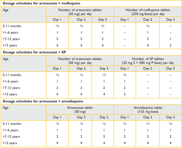
Adapted from: World Health Organization. Manual for the health care of children in humanitarian emergencies, 2008.
Chloroquine-resistant strains of P. falciparum are common throughout many regions of the world. When the proportion of chloroquine-resistant P. falciparum is less than 25%, it may be reasonable to use chloroquine as first-line treatment in less severe patients and assess the response. Failure to respond in 48 to 72 hours indicates infection with a resistant strain. Treat children with chloroquine resistant P. falciparum uncomplicated malaria with quinine sulfate 25 mg/kg/day tid for 3 to 7 days (depending on resistance patterns to quinine) plus one of the following:
- Doxycycline 2,2 mg/kg/day for 7 days (adult dose 100 mg bid for 7 days)
- Tetracycline 25 mg/kg/day qid for 7 days (adult dose 250 mg qid for 7 days)
- Clindamycin 20 mg/kg/day tid for 7 days (adult dose same as for children)
Treat children who may have acquired malaria in Southeast Asia (Thailand) and East Africa with quinine for 7 days, because of the presence of multiple resistant strains in these areas. Additional alternative therapies for resistant P. falciparum include atovaquone-proguanil, mefloquine, halofantrine (associated with heart-related side effects), and artesunate. See appendix for Centers for Disease Control and Prevention (CDC) actual recommended treatment options.
Chloroquine is effective and safe for treating pregnant women. This is important because malaria during pregnancy is more severe and can be fatal. Ideally, use supervised therapy. Observe administration of at least the first dose to be sure that it is not vomited.
If chloroquine phosphate is not available, hydroxychloroquine sulfate is also effective, but in this case 400 mg of hydroxychloroquine are equivalent to 500 mg of chloroquine phosphate.
In several regions mixed infection with species of P. falciparum and P. vivax commonly occurs. If diagnosis of infection is based solely on clinical data, therapy must cover both types of parasites. In the acute phase of an emergency, the detection of a P. falciparum infection is a priority and artemisininbased agents (except artesunate SP) are effective against both organisms.
Supportive management of uncomplicated malaria includes antipyretics, oral rehydration solution (ORS), assessment, and possible referral to a feeding program for malnutrition. Successfully treated patients should improve by 48 hours and be symptom-free by 72 hours. If symptoms persist after 3 days, obtain a new blood smear and consider the possibility of chloroquine-resistant malaria or an alternative cause for the fever.
Treatment of severe and complicated malaria
Assume that all severe, complicated malaria infections are caused by resistant P. falciparum strains unless proven otherwise. Children with severe, complicated malaria can deteriorate rapidly, so initiate treatment with the best available drug and, if possible, arrange a transfer to a hospital for intravenous (IV) therapy (see appendix).
First line (preferred treatment) is Artesunate parentral (IV/IM). In the absence of parenteral form of Artesunate, Artemether IM is acceptable. However, If the child is in shock second choice after artesunate is quinine over artemether.
Quinine is acceptable option but it requires attention to the proper dosage and administration with IV fluids. There is a loading dose and maintenance dose and care needs to be taken to prevent hypoglycemia
- IM artemether Intramuscular (IM) loading dose (3.2 mg/kg) as a single dose on day 1
- Maintenance dose (1.6 mg/kg) IM until the child tolerates oral therapy
- Intravenous (IV) or IM artesunate
- IV loading dose (2.4 mg/kg) over 3 minutes as a single dose on day 1, at 0, 12 and 24 hours
- Maintenance dose (2.4 mg/kg) over 3 minutes, starting on day 2, once a day, until the child tolerates oral therapy
- Rectal artesunate, only if IV or IM routes are not feasible
Administer rectal artesunate 10 mg/kg in a suppository. Repeat the dose if the drug is eliminated within the first hour. Repeat the dose in 24 hours if the patient can not be transferred to the hospital. Artesunate l suppositories remain stable at temperatures of up to 40 degrees and, therefore, require warm, not cold, temperatures for transportation and storage.
After IM or IV therapy, the patient should be switched to oral therapy; the recommended agent in this case is ar temetherlumenfratine (Coartem®) during three days.
If first-line drugs were unavailable, an option is quinine dihydrochloride. A loading dose of 20 mg/kg in 10 mL/kg of 5% dextrose should be administered IV over 4 hours, followed by 10 mg/kg over 4 hours (maximum 1,800 mg/kg) until oral therapy can be started (see Appendix). Blood glucose monitoring for hypoglycemia is recommended every 4 hours after each loading and maintenance infusion. If IV quinine is required for more than 48 hours, maintenance dose should be reduced to 7 mg/base/kg. It is extremely important to bear the infusion volume into account. In order to avoid volume overload due to the IV administration of liquid, the quinine infusion volume should be included in the estimation of daily liquid requirements.
Quinine can be diluted in 5% glucose solution, 10% glucose, 4% glucose, 0.18% saline or 0.9% normal saline. It should be diluted to a total volume of 10 mL/kg (the same volume should be used both for the loading dose and the maintenance dose) and infused over 4 hours. After a minimum of three IV doses of quinine, the patient should be switched to oral therapy. Therapy options by this route include: artemetherlumefantrine (Coartem®) over 3 days, or oral quinine 10 mg base/kg, every 8 hours, until a 7-day course is completed. In areas of multiple resistant malaria, quinine should be combined with oral clindamycin, 5 mg/kg 3 times a day, during 7 days. Mefloquine should be avoided in children that have been in coma, since it increases the risk of neuropsychiatric complications.
A third choice is administering an initial dose of quinine sulfate by oral route or nasogastric tube until IV therapy is available. If the patient vomits, repeat the dose within 30 minutes. If vomits persist, start IM quinidine 10 mg/kg every 4 hours until the patient is transferred to a hospital for IV therapy. At the hospital, treat very severely ill patients with an IV loading dose of quinine gluconate 10 mg/ kg administered over 1 to 2 hours, then with a 0.02 mg/kg/min continuous infusion until oral therapy can be given. If possible, measure hemoglobin, blood sugar, and perform blood and cerebrospinal fluid (CSF) cultures.
Treat children with severe complicated malaria with antibiotics for potential bacteremia or meningitis pending the results of blood and CSF cultures. If possible, monitor the patient for electrocardiographic changes (QT interval, arrhythmias), cinchonism (tinnitus, nausea, headache, and visual changes), and hypoglycemia. Discontinue IV quinidine as soon as the child has improved and switch to oral or nasogastric quinine to complete a 3 or 7 day course (varies by region).
Indications for exchange transfusion vary according to the quality of intensive care facilities and availability and safety of blood products. The theoretical benefits of an exchange transfusion are parasitemia reduction, correction of anemia, improved oxygenation, and enhanced capillary blood flow. It is recommended when children have signs of very severe illness with parasitemia >10%.
The underlying immunopathology of severe dengue infection involves host and viral factors and possibly sequential infections with different virus serotypes.
Supportive treatment of severe, complicated malaria includes antipyretics and oral rehydration solution (ORS). Monitor signs that suggest fluid overload causing pulmonary or cerebral edema. Initial treatment of seizures with at 2-4 mL/kg of dextrose 10% IV or oral 50% dextro, followed by phenobarbital (10 mg/kg IM) if seizures persist.
Seasonal malaria chemoprevention
In seasonal highly endemic areas chemoprevention has been shown to be an effective strategy to reduce malaria epidemics and the incidence of new cases.
7.3. Dengue
Dengue infections occur worldwide but are most prevalent in Southeast Asia, although it has become very prevalent in Central America and the Caribbean and parts of South America. In Southeast Asia, outbreaks of hemorrhagic fever occur cyclically every 4 to 5 years. It is caused by an arbovirus, usually acquired by the bite of Aedes mosquitoes. There are 4 closely related serotypes of dengue virus, all of which can cause severe disease. The underlying immunopathology of dengue infection involves host and viral factors, and possibly sequential infections with different virus serotypes. Dengue infection can have severe clinical manifestations. WHO defines severe dengue by one or more of the following: (i) plasma leakage that may lead to shock (dengue shock) and/or fluid accumulation, with or without respiratory distress, and/or (ii) severe bleeding, and/or (iii) severe organ impairment. After the incubation period, the illness begins abruptly and is followed by the three phases — febrile, critical and recovery (WHO Dengue Handbook 2009)
The underlying immunopathology of severe dengue infection involves host and viral factors and possibly sequential infections with different virus serotypes.
Febrile phase
The acute febrile phase, lasting 2-7 days, usually has a sudden onset of high fever associated with facial flushing, skin erythema, generalized body ache, myalgia, arthralgia and headache. Anorexia, nausea and vomiting are also common. Less frequently sore throat and conjunctival injection can occur. Petechiae, bleeding gums, and or nose bleeds may also occur. There may be an enlarged tender liver during this phase. It is not usually possible to clinically distinguish dengue from other febrile illnesses during this period. However, several laboratory tests are helpful; including a positive tourniquet test, a progressive decrease in the total white cell count and a falling platelet count.
Critical phase
The critical phase begins during a 24-48 hour period when there is an increase in capillary permeability, which is associated with increasing hematocrit levels. This usually occurs after the fever decreases or resolves. Leukopenia and a rapidly decreasing platelet count usually occur just before the increase in capillary permeability. Most patients recover without developing conditions caused by an increase in capillary permeability. In others the severity of the plasma leakage is quite variable. Respiratory distress may result from pleural effusions. Ascites may cause abdominal distention and tenderness. Hemoconcentration is a good indicator of the severity of plasma leakage. When plasma leakage reduces the circulating blood volume signs of shock appear. Prolonged hypoperfusion leads to poor tissue oxygenation, progressive organ failure, metabolic acidosis and disseminated intravascular coagulation (DIC). Signs of shock include a narrowed the pulse pressure (i.e. the difference between the systolic and diastolic pressures ≤20 mm Hg), poor capillary perfusion/delayed capillary refill (>3 seconds), cold extremities, and rapid pulse. Hypotension may be a late manifestation of shock.Consider major bleeding in these cases. Hemorrhage caused by DIC will decrease the hemoconcentration because of blood loss and worsen the shock and organ failure. Massive bleeding may occur without shock in instances when acetylsalicylic acid (aspirin), ibuprofen or corticosteroids have been taken.
Recovery phase
The capillary permeability gradually improves after the 24-48 hour critical phase so that patients begin to stabilize who received appropriate fluid management. There is a reabsorption of fluid and a diuresis. However, bradycardia and electrocardiographic changes are common during this stage.
Dengue Management
In an ambulatory setting where dengue is endemic all patients who have a clinical presentation consistent with the febrile phase of dengue should, if feasible, have an initial CBC to identify neutropenia and decreased platelets and possibly a tourniquet test. They should be followed daily with CBCs until afebrile for 48 hours without decreasing platelets, increasing hematocrits, or other signs of capillary permeability. Warning signs for developing severe dengue include any of the IMCI danger signs, presence of ascites or pleural effusions, increasing hematocrits, signs of shock (narrowed the pulse, poor capillary perfusion/delayed capillary refill, cold extremities, and rapid pulse), severe bleeding, or organ impairment. Make sure that the family understands the importance of returning to the clinic immediately if any of the IMCI danger signs or bleeding develop. Patients who appear to be entering the critical 24-48 phase of dengue with any warning signs should be hospitalized. The decision whether to hospitalize patients without any warning signs and stable CBCs must be individualized based on the degree of illness, access to the hospital and ability of the family to care for the child. It is important to determine if the patient can drink sufficient ORS and fluids. If the patient is unable to drink, start intravenous fluid therapy with 0.9 saline or Ringer’s lactate with or without dextrose at maintenance rate. Patients may be able to take oral fluids after a a few hours of IV therapy. The WHO recommends giving paracetamol for high fever if the patient is uncomfortable. The interval of paracetamol dosing should not be less than six hours. Tepid sponge if the patient still has high fever. Do not give acetylsalicylic acid (aspirin), ibuprofen or other non-steroidal antiinflammatory agents (NSAIDs) as these drugs may aggravate gastritis or bleeding. Acetylsalicylic acid (aspirin) may be associated with Reye’s Syndrome.
All patients with warning signs or severe dengue should be admitted to a hospital with access to intensive care facilities and blood transfusion. Hospital emergency rooms where dengue is endemic should have triage protocols in place to identify severe dengue patients and start fluid management as soon as possible. Triage should classify cases into 3 categories: severe cases needing immediate fluid resuscitation, cases with warning signs who need to be given priority, and non urgent cases. Before beginning IV fluids obtain a reference hematocrit. According to the WHO Dengue handbook avoid administering free water by giving only isotonic solutions such as 0.9% saline, Ringer’s lactate, or Hartmann’s solution. These guidelines are taken directly from the handbook. Start with 5-7 ml/kg/hour for 1-2 hours, then reduce to 3-5 ml/kg/hr for 2-4 hours, and then reduce to 2-3 ml/kg/hr or less according to the clinical response. Reassess the clinical status and repeat the hematocrit. If the hematocrit remains the same or rises only minimally, continue with the same rate (2-3 ml/kg/hr) for another 2-4 hours. If the vital signs are worsening and hematocrit is rising rapidly, increase the rate to 5-10 ml/ kg/hour for 1-2 hours. Reassess the clinical status, repeat the hematocrit and review fluid infusion rates accordingly. Give the minimum intravenous fluid volume required to maintain good perfusion and urine output of about 0.5 ml/kg/hr. Intravenous fluids are usually needed for only 24-48 hours. Reduce intravenous fluids gradually when the rate of plasma leakage decreases towards the end of the critical phase. This is indicated by urine output and/or oral fluid intake that is/are adequate, or hematocrit decreasing below the baseline value in a stable patient. Maintain a detailed fluid balance record and monitor vital signs and peripheral perfusion (1-4 hourly until the patient is out of the critical phase), urine output (4-6 hourly),hematocrit (before and after fluid replacement, then 6-12 hourly), blood glucose, and other organ functions (such as renal profile, liver profile, coagulation profile, as indicated). Care must be taken to avoid excessive fluid administration during the critical phase of capillary permeability to avoid pulmonary edema and congestive heart failure.
The action plan for treating patients with compensated shock in the WHO dengue handbook follows:
- Start intravenous fluid resuscitation with isotonic crystalloid solutions at 5-10 ml/kg/hour over one hour. Then reassess the patient’s condition (vital signs, capillary refill time, hematocrit, urine output). The next steps depend on the situation.
- If the patient’s condition improves, intravenous fluids should be gradually reduced to 5-7 ml/kg/hr for 1-2 hours, then to 3-5 ml/kg/hr for 2-4 hours, then to 2-3 ml/kg/hr, and then further depending on hemodynamic status, which can be maintained for up to 24-48 hours.
- If vital signs are still unstable (i.e. shock persists), check the hematocrit after the first bolus. If the hematocrit increases or is still high (>50%), repeat a second bolus of crystalloid solution at 10–20 ml/kg/hr for one hour. After this second bolus, if there is improvement, reduce the rate to 7–10 ml/kg/hr for 1–2 hours, and then continue to reduce as above. If hematocrit decreases compared to the initial reference hematocrit (<40% in children and adult females, <45% in adult males), this indicates bleeding and the need to cross-match and transfuse blood as soon as possible.
- Further boluses of crystalloid or colloidal solutions may need to be given during the next 24–48 hours.
Avoid fluid overload while ensuring adequate volume replacement. If resources are available, a patient with severe dengue should have an arterial line placed as soon as practical. The reason for this is that in shock states, estimation of blood pressure using a cuff is commonly inaccurate. The use of an indwelling arterial catheter allows for continuous and reproducible blood pressure measurements and frequent blood sampling on which decisions regarding therapy can be based. Monitoring of ECG and pulse oximetry should be available in the intensive care unit. Urine output should be checked regularly (hourly till the patient is out of shock, then 1-2 hourly). A continuous bladder catheter enables close monitoring of urine output. An acceptable urine output would be about 0.5 ml/kg/ hour. Hematocrit should be monitored (before and after fluid boluses until stable, then 4-6 hourly). In addition, there should be monitoring of arterial or venous blood gases, lactate, total carbon dioxide/bicarbonate (every 30 minutes to one hour until stable, then as indicated), blood glucose (before fluid resuscitation and repeat as indicated), and other organ functions (such as renal profile, liver profile, coagulation profile, before resuscitation and as indicated).
Severe bleeding can be recognized by:
- persistent and/or severe overt bleeding in the presence of unstable haemodynamic status, regardless of the hematocrit level;
- a decrease resuscitation in hematocrit haemodynamic together status;
- Blood transfusion is life-saving and should be given as soon as severe bleeding is suspected or recognized.
The action plan for the treatment of hemorrhagic complications is as follows:
- Give 5-10 ml/kg of fresh-packed red cells or 10-20 ml/kg of fresh whole blood at an appropriate rate and observe the clinical response. It is important that fresh whole blood or fresh red cells are given. Oxygen delivery at tissue level is optimal with high levels of 2,3 di-phosphoglycerate (2,3 DPG). Stored blood loses 2,3 DPG, low levels of which impede the oxygen-releasing capacity of haemoglobin, resulting in functional tissue hypoxia. A good clinical response includes improving hemodynamic status and acid-base balance.
- Consider repeating the blood transfusion if there is further blood loss or no appropriate rise in hematocrit after blood transfusion. There is little evidence to support the practice of transfusing platelet concentrates and/or fresh-frozen plasma for severe bleeding.
7.4. Chikungunya
Chikungunya virus, which like dengue is transmitted to people by Aedes species mosquitoes, causes a febrile illness that initially may be indistinguishable from dengue. Infection is characterized by fever, myalgia, and lethargy. Additional symptoms may include a maculopapular rash, nausea, vomiting, and headache. The infection may progress to debilitating bilateral polyarthralgia and, in some cases, arthritis. The virus is endemic in many countries Africa, Asia, Europe, and the Indian and Pacific Oceans. In late 2013, chikungunya virus was found for the first time in the Americas on islands in the Caribbean and it has now become endemic throughout the Caribbean and tropical areas of Latin America. Chikungunya infection is rarely fatal compared to dengue. However, Chikungunya virus, which like dengue is transmitted to people by Aedes species mosquitoes, causes a febrile illness that initially may be indistinguishable from dengue. Infection is characterized by fever, myalgia, and lethargy. Additional symptoms may include a maculopapular rash, nausea, vomiting, and headache. The infection may progress to debilitating bilateral polyarthralgia and, in some cases, arthritis. The virus is endemic in many countries Africa, Asia, Europe, and the Indian and Pacific Oceans. In late 2013, chikungunya virus was found for the first time in the Americas on islands in the Caribbean and it has now become endemic throughout the Caribbean and tropical areas of Latin America. Chikungunya infection is rarely fatal compared to dengue. However, roidal anti-inflammatory drugs (e.g., aspirin and ibuprofen) should be avoided and fever and pain should be managed with acetaminophen. Prevention by avoiding mosquito bites remains the most effective strategy for both dengue and chikunguya.
7.5. Ebola Virus Disease
The 2014 Ebola Virus Disease (EVD) outbreak in West Africa underscored the importance of preparedness, pre-planned response, and mitigation of infectious and contagious diseases. EVD demonstrated that poorly-resourced African countries, with unprepared, fragile, and strained healthcare systems, were particularly susceptible and crippled by the outbreak. Repeat outbreaks and a possible global pandemic is still a reality.
Epidemiology
EVD was discovered in the former republics of Zaire and Congo In 1976 by World Health Organization scientists. There have been approximately 20 outbreaks since its discovery, mostly clustered in sparsely populated towns and villages in Central Africa.2 The 2014 EVD West African outbreak occurred in densely populated urban areas in West Africa. Ultimately, EVD caused over 27,000 cases and claimed over 11,000 lives, and nearly cascaded into a global pandemic.1 The overall social, economic, and public health toll were incalculable. It is estimated that the nations of Liberia, Guinea, and Sierra Leone will take approximately 10 years to return to their pre-Ebola state.
Pathogenesis and Transmission
Five species of virus, of the family Filoviridae, have been identified that cause Ebola Virus Disease; Zaire, Bundibugyo and Sudan ebolaviruses have been associated with outbreaks in Africa. Ebolavirus is transmitted through bodily fluids. The natural host of ebolavirus is wild animals, including fruit bats. Traditional practices, i.e. the consumption of undercooked infected bush meat, is believed to be a frequent cause of EVD in humans. Human-to-human transmission is through direct contact in which infected bodily fluids transmit ebolavirus. The mean incubation period is 5 days (range: 2-21 days).4 As the viral burden increases in a human, the human becomes more clinically symptomatic, and more infectious; an infected person is most contagious upon death, and corpses can harbor live ebolavirus for weeks. It should be emphasized that humans are not infectious until they develop symptoms. This places healthcare workers at particularly high risk for EVD during routine patient care. It also places family members, caretakers off ill, and handlers of corpses at highest risk of catching EVD.
Clinical Syndrome
EVD is characterized by a non-specific or constitutional illness, most similar to a viral illness. Its clinical syndrome includes fever (87.1%), fatigue (76.4%), loss of appetite (64.5%), vomiting (67.6%), diarrhea (65.6%), headache (53.4%), and abdominal pain (44.3%).3 EVD symptoms overlap considerably with influenza and malaria, and may be confused with typhoid fever and meningitis. It should be noted that hemorrhage occurs in less than 20% of cases, but is more likely to predict death. Bleeding, if present, occurs mostly in GI and respiratory mucosa, and at the site of intravenous punctures. Hence, it may cause hemoptysis and hematochezia.
Screening and Quarantine
Because Ebola virus is spread through contact with the body fluids of infected, symptomatic patients, transmission can be stopped by a combination of early diagnosis, contact tracing, patient isolation and care, infection control, and safe burial.3 In healthcare settings, it is critical that a form of non-contact screening be performed of all patients arriving at the healthcare facility. In the 2014 West African outbreak, this was best accomplished using infrared no contact temperature sensors and screening questions, performed with a minimum separation of 3-feet between healthcare provider and patient. A positive screen for EVD includes fever, contact with a person with EVD, and/or travel to an endemic area. Ebolavirus may be confirmed by polymerized chain reaction (PCR) or enzyme-linked immunosorbent assay (ELISA) testing. Patients who screen positive should be quarantined to assess development of clinical symptoms, and/or repeat confirmatory testing.\( ^{4} \)
Treatment
As of early 2015, no specific cure, or vaccine exists for ebolavirus infection in humans. The standard and hallmark of care is supportive therapy. This includes basic acute and resuscitative care, including: hydration, electrolyte repletion (if vomiting or diarrhea), fever control, and physical safety for altered patients. It is critical that healthcare workers adhere to strict personal protective equipment guidelines set forth by international health agencies. Further, they should minimize performance of any potentially “risky” procedures e.g. intravenous insertions or blood draws, or avoid spreading ebolavirus through nebulizer treatments, positive pressure ventilation, or leakage of infected bodily fluids.3 Of note, healthcare workers faced disproportionately higher rates of infection and death than the general population, speaking to their particular vulnerability, need for education and personal protection. \( ^{1} \)\( ^{,} \)\( ^{4} \)
Healthcare System Management of Ebola
EVD directly threatens patients, healthcare workers, healthcare systems, communities, and nations. It is a considerable public health threat, and by all accounts, outbreaks of EVD in low-resource settings are a healthcare disaster in their own right. Healthcare systems need to prepare and plan by developing disease surveillance systems to monitor and contain small outbreaks before they spread. Facilities need to have pre-planned procedures and researched drills for how to secure additional resources, use personal protective equipment, and how to re-configure their physical spaces (to quarantine and observe suspected cases, and separately treat confirmed or probable cases, while protecting healthcare workers and the general public). Outbreak containment measures, such as mandatory safe burials by specialist burial teams, are essential.4 Communication, through sustained community engagement, also proved to be a successful method of disseminating public health information during the West Africa outbreak. \( ^{4} \)
References:
- World Health Organization. Ebola Data and Statistics: Situation Summary http://apps.who.int/gho/data/ node.ebola-sitrep.ebola-summary?lang=en Accessed June-25-2015.
- Centers for Disease Control and Prevention Ebola outbreaks 2000-2014. 2014.
- WHO Ebola Response Team. N Engl J Med 2014; 371:1481-1495, October 16, 2014DOI: 10.1056/ NEJMoa1411100
- World Health Organization. Fact Sheet, No 103: Ebola Virus Disease. http://www.who.int/mediacentre/factsheets/fs103/en/ Access June-25-2015.
7.6. HIV
Impact of Pediatric HIV
In 2013, 3.2 million children were estimated to be living with HIV, 90% of them in sub-Saharan Africa.1 Despite substantial progress in preventing mother to child transmission, 240,000 children less than 15 years of age are HIV infected every year.1 Only 1 out of 4 children living with HIV have access to life-saving anti-retroviral treatment (ART). Amongst children born with HIV, 50% will die by 2 years of age without treatment.2 Keys to caring for children with HIV include 1) recognizing children and risk for HIV infection and diagnosing them early, 2) treating all children under 5 years with HIV and all those eligible for treatment >5 years, 3) managing co-infections and opportunistic infections, and 4) ensuring excellent adherence by avoiding missed doses and treatment interruptions.
Recognition and Diagnosis of HIV
Key concepts:
- Child is under 18 months: HIV infection is confirmed if virological test (PCR) is positive
- Child is over 18 months: HIV infection is confirmed if two diff erent serological (antibody) tests are positive
Recognition of children at risk for HIV infection is key to identifying these children early and initiating life-saving therapy rapidly. The majority of children are infected through mother to child transmission. Therefore, diagnosing HIV in women of childbearing age is critical to not only caring for women with HIV but preventing new pediatric infections. ALL pregnant women should undergo voluntary HIV testing and receive prophylaxis and treatment as recommended by in-country guidelines. Currently, WHO recommends combination anti-retroviral therapy for all pregnant and breastfeeding women in areas with high HIV burden and suggests ART be continued throughout life. \( ^{3} \)
Children born to women with known HIV infection or unknown status must be prioritized for HIV testing. Children under 18 months of age may carry maternal HIV antibodies in their blood indicating HIV exposure but not confirming HIV infection. Therefore, regular HIV antibody testing (such as rapid tests) may be used to confirm HIV exposure in those <18 months of age and HIV infection in those >18 months of age. If a child is HIV exposed (positive antibody test or mother with known HIV infection) virological testing using polymerase chain reaction (PCR) is required through a specialized laboratory.
Treatment of HIV
Key concepts:
- All children less than 5 years who are HIV infected should be initiated on ART irrespective of CD4 count or clinical stage.
- If a child has any general danger sign or a severe classification, he or she needs URGENT REFERRAL. ART initiation is not urgent, and the child should be stabilized first.
- Early infant diagnosis in high prevalence areas must be a priority
- PMTCT should be reinforced
WHO eligibility criteria for ART initiation in children\( ^{3} \)
- ART should be initiated in all children infected with HIV below five years of age, regardless of WHO clinical stage or CD4 cell count
- ART should be initiated in all HIV infected children five years of age and older with CD4 cell count ≤500 cells/mm3 , regardless of WHO clinical stage
- ART should be initiated in all children infected with HIV with severe or advanced symptomatic disease (WHO clinical stage 3 or 4) regardless of age and CD4 cell count
- ART should be initiated in any child younger than 18 months of age who has been given a presumptive clinical diagnosis of HIV infection
Below are IMCI simplified approach for starting ART in children. Whenever possible, also refer to in-country guidelines. \( ^{4} \)
Steps in initiating ART for children
- Step 1 DECIDE IF THE CHILD HAS CONFIRMED HIV INFECTION
- If HIV is confirmed (see abo ve) and child is in stable condition, go to STEP 2.
- If HIV is confirmed (see abo ve) and child is in stable condition, go to STEP 2.
- Step 2 DECIDE IF CAREGIVER IS ABLE TO GIVE ART
- Check that the caregiver is willing and able to give ART. Support caregiver to disclose child’s HIV status to another adult who can assist in giving ART.
- Caregiver able to give ART: GO TO STEP 3
- Caregiver not able: classify as CONFIRMED HIV INFECTION but NOT ON ART. Counsel and support the caregiver. Follow-up regularly. Move to the step 3 once the caregiver is willing and able to give ART
- Step 3 DECIDE IF ART CAN BE INITIATED IN YOUR FACILITY
- If child is less than 3 kg or has TB, Refer for ART initiation.
- If child weighs 3 kg or more and does not have TB, GO TO STEP 4
- Step 4 RECORD BASELINE INFORMATION ON THE CHILD’S HIV TREATMENT CARD
- Record the following information:
- Weight and height
- Pallor if present
- Feeding problem if present
- Laboratory results (if available): Hb, viral load, CD4 count and percentage. Send for any laboratory tests that are required. Do not wait for results.
- GO TO STEP 5
- Step 5 START ON ART, COTRIMOXAZOLE PROPHYLAXIS AND ROUTINE TREATMENTS
- Child up to 3 years: ABC or AZT +3TC+ LPV/R or recommended firstline regimen
- Child 3 years or older: ABC + 3TC + EFV, or recommended first-line r egimen.
- Give co-trimoxazole prophylaxis
- Give other routine treatments, including Vitamin A and immunizations
- Follow-up regularly as per national guidelines
- Step 1 Decide if the Child has Confirmed HIV Infection
- Step 2 Decide if Caregiver is able to Give ART
- Step 3 Decide if ART can be Initiated in YourFacility/Location
Treatment of Co-Infections and Opportunistic Infections
Children exposed to and living with HIV are generally at higher risk of the common childhood illnesses including diarrhea, pneumonia, malnutrition, and TB. Early recognition and treatment as per IMCI and National guidelines are critical. Prevention is equally important—all HIV exposed and infected children should receive routine immunizations on schedule including live vaccines unless they are severely immunosuppressed. Additionally, in most developing country settings, prophylaxis of PCP is recommended for all exposed and infected children using co-trimaxozole. More recently, prophylaxis for TB has been recommended in countries with high TB burden using isoniazid. Refer to in-country guidelines.
HIV Management in Disaster or Displacement Scenarios
Initial management of HIV-infected children should include documentation of last CD4 and viral load if known and ART regimen if child taking ART. Children who had their antiretroviral therapy and/or prophylaxis or treatment for OIs interrupted by disaster-related displacement should restart these medications as soon as possible. There is no need to start antiretroviral therapy immediately in those HIV-infected patients who were not receiving antiretroviral medications before the disaster.\( ^{5} \)\( ^{,} \)\( ^{6} \)
It is important to determine the current antiretroviral drugs the child is taking. If this is unknown and records are unavailable, consider consultation with an HIV specialist or starting first line treatment as recommended by WHO or national guidelines. All antiretroviral drugs should be restarted at the same time except if the child was receiving nevirapine as part of a regimen and has not taken nevirapine for more than 7 days. If this occurs, nevirapine should be restarted with a 2-week “lead-in” period, giving half the daily dose once daily for 14 days and then standard twice-daily dosing. Of note, some children may be taking liquid formulations, some of which may need to be refrigerated. Pay special attention to ensure proper dosing based on weight-based dosing recommendations.
Treatment of acute infections
HIV-exposed and infected children may be more vulnerable to the usual acute infections seen in disasters such as diarrhea, acute respiratory infections, measles etc. Treatment for acute illnesses remains the same in children with and without HIV.
Prevention of OIs
Prevention of opportunistic infections is very important amongst HIV-infected and exposed children. Routine prophylaxis for OIs with co-trimoxazole as recommended above should be maintained if at all possible following a disaster. Of note, cotrimoxazole is commonly used for routine treatment of ARI’s which may lead to drug shortages and difficulty maintaining drug stock for both acute treatment of illness and prophylaxis. Additional precautions to prevent TB should be emphasized for individuals with HIV, especially children under 5.
Immunizations
HIV-exposed and infected children should receive all routine immunizations on schedule including live vaccines unless they have symptoms of severe immunosuppression or are severely ill. HIV-infected children are particularly susceptible to complications of measles and receive measles vaccines during should disaster situations or displacement conditions unless severely immunosuppressed.
8. Other Cases That Require Attention at the Scene of the Disaster
Objectives
- Distinguish other clinical entities that can present at the scene of the disaster, such as tuberculosis.
- Consider meningitis in emergency settings and assess the clinical findings.
Tuberculosis
Even though tuberculosis (TB) is the leading infectious cause of death in some parts of the developing world, TB treatment and control programs are not part of an emergency relief response. TB is a chronic infection and effective treatment is very resource-intensive. Treatment programs need to include resources to identify and monitor true cases by sputum smears exam, a stable population for at least 6 months (to complete shortcourse therapy), enough available drugs to treat all cases, and enough personnel to supervise all therapy in the first 2 to 3 months. Administration of anti-TB drugs to persons who will not adhere to or complete treatment is likely to contribute to drug resistance in the community.
Even though tuberculosis (TB) is the leading infectious cause of death in some parts of the developing world, TB treatment and control programs are not part of an emergency relief response.
Meningitis
Meningitis is the inflammation of the membranes (meninges) that surround the brain and spinal cord. Encephalitis is the inflammation of the cerebral Meningoencephalitis cortex. involves both the meninges and the cerebral cortex.
Meningitis may be due to viral, bacterial, or fungal infections. Approximately two thirds of diagnosed cases are viral and one third are bacterial. The most common viral infections are caused by enteroviruses and herpes simplex virus.
The most common bacterial pathogens that cause meningitis during the first 3 months of life include group B Streptococcus (GBS), Escherichia coli, Listeria monocytogenes, enterococci, Staphylococcus aureus, and gram-negative enteric organisms. The viral pathogens in this age group are herpes simplex virus, enterovirus, and cytomegalovirus.
Pathogens infecting infants older than 3 months of age and children are most often S. pneumoniae, Haemophilus influenzae type b (Hib) and Neisseria meningitidis. Other organisms such as M. tuberculosis, Salmonella, and Mycoplasma pneumoniae are rare.
The frequency of Haemophilus infection has dramatically decreased with immunization. However, in areas of the world where conjugate Hib vaccine is not administered, this organism remains a common cause of meningitis. Viral pathogens most prevalent in this arbo age group include enterovirus, virus, herpes simplex virus, herpes virus, influenza, and Epstein-Barr virus.
Clinical findings of meningitis
Look for changes in mental status and level of activity, including irritability, changes in feeding and sleeping patterns, unresponsiveness, and seizures.
Check for signs of meningeal irritation: nuchal rigidity, bulging fontanelle, paradoxical irritability, and Brudzinski and Kernig signs.
Evaluate hydration status and signs of shock, such as mottled skin, slow capillary refill, increased pulse, and decreased blood pressure. Perform a neurologic examination and document focal neurologic signs, paresis, or ataxia. Measure the head circumference and look for exanthem, purpura or petechiae, or soft-tissue, bone, or joint infections.
Signs associated central nervous system complications include focal neurologic findings, prolonged seizures, persistent changes in mental status, enlarging head circumferences, or ataxia. Complications include subdural effusion or empyema, cerebral edema, cerebral abscess, cerebral infarction, or hydrocephalus.
Treatment of meningitis
Suspected cases of severe sepsis or meningitis need to be treated promptly with the best available drugs. Report such cases to health authorities and make attempts to obtain appropriate samples for identification of the causative agent.
Identification of Neisseria meningitidis is particularly important because of its epidemic potential and the fact that a reasonably effective vaccine is available.
During confirmed N. meningitidis outbreaks, chemopr implement vaccination and ophylaxis of household contacts.
N. meningitidis remains susceptible to penicillin all over the world. A long-acting suspension of chloramphenicol in oil, called tifomycin, could be an alternative to penicillin. When other antibiotics are available, the initial antibiotic therapy depends on the age of the patient. Treat newborn infants with ampicillin and an aminoglycoside (gentamicin) or cefotaxime. Ampicillin is needed to cover Listeria and enterococci. Treat infants 1 to 3 months of age with ampicillin and ceftriaxone, or cefotaxime to cover enterococcus, Listeria, and H. influenzae. Treat older children with vancomycin and ceftriaxone if the rate of penicillin-resistant S. pneumoniae in the area is high.
When non susceptible organisms are identified, consider the recommended high doses of cefotaxime and ceftriaxone, and add rifampin when the minimum inhibitory concentration (MIC) of the nonsusceptible pneumococci is >2.0 µg/ mL. If possible, obtain serum creatinine levels before giving vancomycin and repeat weekly during treatment, because vancomycin excretion depends on glomerular filtration.
When non susceptible organisms are identified, consider the recommended high doses of cefotaxime and ceftriaxone, and add rifampin when the minimum inhibitory concentration (MIC) of the nonsusceptible pneumococci is >2.0 µg/ mL. If possible, obtain serum creatinine levels before giving vancomycin and repeat weekly during treatment, because vancomycin excretion depends on glomerular filtration.
When using aminoglycosides or chloramphenicol, monitor blood levels if possible (therapeutic levels for gentamicin or tobramycin are 4 to 8 µg/mL; for kanamycin or amikacin, 15 to 25 µg/mL). Adequate blood chloramphenicol levels can be achieved with oral administration. Whenever possible, avoid administering aminoglycosides in patients with renal disease and chloramphenicol in patients with hepatic dysfunction.
9. Vaccination in Disaster Situations
Objectives
- Acknowledge the importance of measles immunization in a disaster situation.
- Recognize the characteristics of tetanusprone injuries and wounds.
- Describe specific situations that require the use of other vaccines.
The only vaccine that must be routinely administered during immediate emergency relief efforts is measles. A routine immunization program for other vaccines should only be considered if the population is expected to stay in the area for longer than 3 months, if it is possible to keep appropriate records, and if other assistance efforts are not disrupted or compromised by the activities needed for vaccination.
Tetanus
Tetanus immunization is not routinely recommended in disaster situations, but if the vaccine is available, it is reasonable to apply it prophylactically to individuals who have tetanus-prone wounds if the time of the last tetanus immunization is unknown or greater than 5 years, or when the child has not received the primary 3-dose vaccination series. The characteristics of tetanus-prone wounds are a wound that was first cleaned more than 6 hours after its occurrence; irregular wounds; wounds from bullets, crushing, burns, or frostbite; and presence of devitalized wound contaminants.
Specific situations requiring prophylaxis
Pertussis
The vaccine. It is well established that pertussis vaccine provides clinical protection after exposure to the disease in most people. The effectiveness of the vaccine with a regimen of 3 or more doses is around 80% to 90%. Appropriately immunized children who acquire the disease have milder symptoms and fewer complications.
Management of outbreaks. When an increase in the number of cases is suspected, mass immunization is a priority in children under 7 years old. If disease rates are higher among children over 7 years old and adolescents, use of acellular vaccines may be considered.
Household contacts—vaccination. Household contacts and other close contacts of patients under 7 years old who have had at least 4 previous doses of diphtheriatetanus-pertussis vaccine (DTP or DTaP) must receive a booster injection of DTP or DTaP, unless they have received a dose within the past 3 years. Children under 7 years old who have not been immunized or who have previously received less than 4 doses must start or continue their vaccination regimen according to the national program. A fourth dose must be administered to children who received their third dose 6 or more months before exposure.
Chemoprophylaxis. All household contacts and other close contacts, regardless of their age or immune status, should receive erythromycin (40-50 mg/kg/day orally, divided in 4 doses), for 14 days because immunity after vaccination is not total and infection may not be prevented. It has been proven that erythromycin eliminates the carrier state and is effective in limiting secondary spread. For patients who are intolerant to erythromycin, clarithromycin (15 mg/kg/day orally divided in 2 doses, for 1 week) may be administered; other options are azithromycin and trimethoprim-sulfamethoxazole.
Diphtheria
The vaccine. In diphtheria, as in tetanus, immunity relies only on the presence in blood and interstitial fluids of antitoxin IgG antibodies with titers ≥0.01 IU/mL. These antibodies work locally, where the toxin is released by the bacteria, and in blood against the toxin that reaches the circulation. After primary immunization with 3 doses of toxoid, antitoxin titers above 0.01 IU/mL can be found for 5 or more years, and after one or more booster injections they persist for 10 years. In clinical practice, vaccination has shown an efficacy rate above 99%.
Management of outbreaks. When cases of diphtheria are suspected, mass vaccination is indicated, taking into account the rates of incidence by age groups.
Household contacts—vaccination. Asymptomatic contacts whose immunization regimen is complete and who have received their last dose more than 5 years ago must receive a DTP or dT booster according to their age. Close asymptomatic contacts whose immunization regimen is incomplete (<3 doses of diphtheric toxoid) or whose immunization status is unknown must receive a dose and complete the schedule.
Chemoprophylaxis. Whatever their immunization status, close contacts must be kept under surveillance for 7 days to detect any evidence of the disease, have cultures taken for Corynebacterium diphtheriae, and receive antimicrobial prophylaxis with oral erythromycin (40-50 mg/ kg/day for 7 days, with a maximum of 2 g/day) or a single intramuscular (IM) dose of penicillin G benzathine (600,000 IU for those <30 kg and 1.2 million units for older children and adults). Obtain new throat cultures in contacts identified as carriers within 2 weeks after completion of treatment.
Meningococcal disease
Few infectious diseases cause as much concern among the general population and health workers as meningococcal infection. The estimated attack rate for household contacts is 4 cases per 1,000 exposed persons. This is 500 to 800 times higher than rates in the general population.
Chemoprophylaxis is indicated for those individuals who meet the criteria for close contacts. The goal is to eradicate N. meningitidis carriers and prevent the occurrence of secondary cases.
- Close contacts: household members, attendees at child care centers, nursery schools, schools, universities, and members of closed communities that are in contact with any individual with mening ococcal disease for more than 4 hours daily, 5 days a week; any other person directly exposed to oral secretions of the patient (e.g., sharing tableware, drinks, kisses; sneezing or coughing).
- Secondary case: any case occurring in a close contact 24 hours or more after onset of the disease in the primary case. Because there is a high rate of secondary disease during the 5 days following contact, give chemoprophylaxis within the first 24 hours. It is not indicated beyond 14 days. A nasopharyngeal culture to determine the need for chemoprophylaxis is not warranted. If the patient was treated with third generation cephalosporins, chemoprophylaxis before discharge is not needed. Rifampin is the first choice agent for chemoprophylaxis in children, but there are alternatives for adults (Table 4). Chemoprophylaxis is indicated for household members and contacts (Box 5). Monitor exposed individuals and assess if they have a febrile disease.
Table 4. Recommended Agents for Chemoprophylaxis
| Rifampin | |
|---|---|
| Newborns | 5 mg/kg/dose |
| Children | 10 mg/kg/dose |
| Adults | 600 mg/dose |
| Duration | Every 12 h for 2 days |
| Ceftriaxone | |
|---|---|
| ≤12 years | 125 mg IM |
| >12 years | 250 mg IM |
| Pregnant women | 250 mg IM |
| Duration | Single dose |
| Ciprofloxacin | |
|---|---|
| Age group | ≥18 years |
| Dose | 500 mg orally |
| Duration | Single dose |
IM: Intramuscular.
BOX 5: Indications for chemoprophylaxis
Contacts who should receive chemoprophylaxis
- Household members
- Individuals who often sleep or eat with the patient, and meet the definition of contact
- Contacts in child care centers and nursery schools (including staff members) for more than 4 hours during 5 days of the previous week
- Individuals who have been directly exposed to the patient’s secretions through kissing or sharing food, drinks, toothbrushes, etc.
- Individuals administering mouth-to-mouth resuscitation
- Individuals who experience unprotected contact during endotracheal intubation in the 7 days prior to the onset of the disease
Situations where chemoprophylaxis is NOT indicated:
- Casual contact: no direct exposure to the patient’s oral secretions (classmates or coworkers)
- Indirect contact: no contact with the patient, only with his/her contact
- Health care workers with no direct exposure to the patient’s oral secretions
The vaccine: immunogenicity and effectiveness. With unconjugated polysaccharide vaccines, protection is achieved 7 to 10 days after immunization. Bivalent A + C vaccine is safe and effective (85% to 90%) in children older than 2 years old and in adults. The A component induces an immune response from 3 months of age on, with a seroconversion rate of 88% after the second dose, applied in children between 7 and 12 months old.
Management of outbreaks. An outbreak of meningococcal disease is defined when the attack rate is higher than 10 cases in 100,000 persons, in a specific area, with an epidemiologic relation among cases, and with a predominating serogroup. With active epidemiologic surveillance, an outbreak is also considered when the incidence rate by age is doubled.
Where can outbreaks occur? Outbreaks can occur in an institution or an organization. In this case, an outbreak is defined by 3 or more confirmed, presumptive, or probable cases occurring in a period of 3 months or less within the same institution or organization, but without close contacts (e.g., schools, universities, military organizations, jails).
Community outbreaks are defined by 3 or more confirmed, presumptive, or probable cases that occur in 3 months or less among people who live in the same area and are not close contacts (e.g., small towns, cities, countries).
9.1. Guidelines for Evaluation and Management of a Meningococcal Disease Outbreak
1. Reinforcement of active surveillance
In areas where surveillance for meningococcal disease is passive, case reports may be incomplete or delayed. When an outbreak is suspected, alert public health authorities and request immediate report of new cases.
2. Case detection and bacteriologic confirmation
Establish the diagnosis of meningococcal disease considering confirmed, presumptive, or probable cases:
- Confirmed case: isolation of N. meningitides from a usually sterile site (blood, CSF) in an individual with clinically consistent findings.
- Presumptive case: observation of Gram-negative diplococci in any usually sterile site, with negative cultures and symptoms of disease.
- Probable case: positive antigen test for N. meningitidis (latex agglutination test, immunoelectrophoresis), with negative cultures and consistent symptoms.
Information about serogroup is essential. Laboratories not performing this test routinely should forward the sample to referral laboratories of higher complexity to identify the serogroup. If possible, investigate N. meningitidis subtype by pulsedfield gel electrophoresis or multilocus enzyme electrophoresis to determine if the strains of a group of cases are interrelated and whether they represent an outbreak.
Co-primary case:
Case occurring in a close contact within 24 hours of the onset of the disease in the primary case
3. Appropriate treatment of patients, according to management guidelines
4. Chemoprophylaxis and careful observation of contacts
Chemoprophylaxis and careful observation are recommended for close contacts. Chemoprophylaxis for individuals who are not close contacts is ineffective in preventing community outbreaks; therefore, it is not recommended. Exposed individuals must be carefully monitored and evaluated in case of any febrile illness.
5. Investigation of relationships between cases
In addition to demographics, obtain the following information for each affected individual: history of close contact with another primary case; participation in social activities or sports; attendance at child care centers, kindergartens, schools, universities, or clubs. This information will help identify cases as co-primary or secondary, reveal relationships between cases, and define the population at risk.
6. Assessment of the relationship of the suspected outbreak with the community or with an institution or organization
7. Definition of at-risk population
In outbreaks related to an institution or organization, cases are linked with a shared affiliation, such as attending the same day care center, kindergarten, school, or university or belonging to the same sports team. In such cases, the population at risk is everyone in those places. On the other hand, in community outbreaks patients do not share an affiliation, only a geographically defined location, such as a neighborhood, small town, city, or country. The risk group includes every individual living in those places.
8. Estimation of attack rate
Attack rate can be estimated by the following formula:
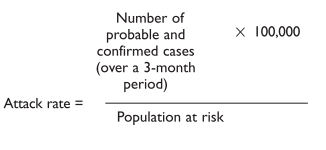
With a global attack rate higher than 10 cases in 100,000, consider vaccination of at-risk population. Consider the incidence rates by age groups. If the incidence rate doubles in a population with adequate epidemiological sur veillance, immunization may be considered.
9. Selection of the target group for vaccination
Consider the guidelines from public health authorities regarding the serogroup involved and the age group affected. In that case, it is necessary to have adequate vaccine supplies.
10. Infections in Infants 0 to 2 Months of Age
OBJECTIVES
- Identify and establish the treatment for sick infants 0 to 2 months of age.
Assessment of the sick infant 0 to 2 months of age
As mentioned previously, newborn and infants under 2 months of age are very vulnerable to infections, with high morbidity and mortality rates associated with very severe clinical conditions including sepsis, meningitis, and pneumonia. Thus, if an infant under 2 months is suspected of having a severe neonatal illness or a possible severe bacterial infection, there is no time to lose in laboratory studies. It is extremely important to start antibiotic therapy immediately and to refer the patient to a hospital if needed resources are not available.
Infants weighing less than 2,000 g who are brought to the primary health care facility with a possible infection should be referred to a hospital for specialized treatment, regardless of the severity of the condition, because they are more vulnerable due to their immaturity. The assessment of the infant 0 to 2 months ld should include the following questions:
- What is your baby doing?
- Is he/she feeding well or poorly?
- Has he/she vomited / Is he vomiting all he eats?
- Has he/she had diarrhea?
- Has he/she difficulty breathing?
- Has he/she had fever or hypothermia?
- Has he/she had seizures or shivering?
Infants 0 to 2 months old who need to be transferred to a hospital more than 5 hours away should receive an intramuscular dose of an adequate antibiotic.
In addition, look for clinical signs that indicate the severity of the illness, from subtle signs such as “he doesn’t look good” to neurologic signs (e.g., seizures) or difficult breathing. Assessment of body temperature, hydration status, capillary refill, and fontanelle characteristics are also important, as well as looking for other problems (congenital anomalies, surgical disorders). Figure 3 shows the algorithm used by the Integrated Management of Chilhood Illness (IMCI) for assessment of sick infants 0 to 2 months of age. The IMCI strategy includes the following danger signs: not feeding, convulsions, fast breathing (more than 60 breaths per minute) severe chest indrawing, fever or low temperature, and lack of movement.
Young children having any of these signs of very severe disease is classified as Pink and should be urgently referred to the hospital with a first antibiotic dose and treatment to prevent low blood sugar. A young child with signs of umbilical infection (redness and or purulent discharge) or skin pustules is classified as yellow having a local bacterial infection and treated with an appropriate antibiotic.
Classification of the infant 0 to 2 months of age with severe illness or possible severe bacterial infection
It is difficult to distinguish between a very severe illness and a severe infection, such as sepsis or meningitis, since clinical findings are usually similar. For this reason, the classification gives both possibilities.
If the infant is suffering from a local but extensive bacterial infection, he or she should also be classified as having a possible severe bacterial infection because the local infection can disseminate and result in sepsis, due to the immaturity of the immune system. He or she needs to be referred urgently to a specialized hospital to receive different kinds of treatments, such as oxygen or parenteral antibiotics. Before transfer, administer the first dose of the adequate antibiotic. Transfer according to the guidelines for stabilization and transport. Counsel the mother or caregiver in order to clarify possible doubts and provide support.
Treatment of infants 0 to 2 months of age with infection
Infants 0 to 2 months old who need to be transferred to a hospital more than 5 hours away should receive an intramuscular (IM) dose of an adequate antibiotic.
Possible antibiotic combinations include:
- gentamicin + ampicillin
- gentamicin + G penicillin procaine
Avoid oral feeding if the infant presents with altered consciousness or difficult breathing, and administer a 5% dextrose solution through nasogastric tube to prevent hypoglycemia.
If an infant less than 2 months is suspected of having a severe neonatal illness or a possible severe bacterial infection, there is no time to lose in laboratory studies.
If there is no incubator available for the transfer, the “mother kangaroo” technique is advisable in order to prevent hypothermia. If available, also administer supplemental oxygen during the transfer to prevent hypoxemia.
Infants 0 to 2 months of age with a local bacterial infection should receive an adequate oral antibiotic as well as topical antibiotic therapy according to the site of infection.
11. Summary
The morbility and mortality associated with infectious diseases are very high in developing countries. During acute humanitarian emergencies, morbility and mortality increase significantly. Deterioration of the nutritional status associated with such situations increases the risk of infectious diseases among the affected children.
The IMCI strategy, designed for primary care management of children and based on a number of clinical signs at presentation, is an ideal tool for the effective management of people affected by disasters, particularly in situations with limited resources, both material and human. This tool allows a quick and simple distinction between children who require referral to the hospital and those with less severe illness that can be managed in a less complex setting.
Measles, acute respiratory infections, malaria, dengue, and acute diarrhea are the infections that cause more concern in an emergency setting. Take also into consideration sepsis and meningitis. It is important to recognize these diseases as early as possible in order to give appropriate therapy and prevent a possible outbreak among people displaced by a disaster.
12. Suggested Reading
- Black RE. Persistent diarrhea in children in developing countries. Pediatr Infect Dis J 1993;12:751-761.
- Black RE, Morris SS, Bryce J. Where and why are 10 million children dying every year? Lancet 2003;361:2226-2234.
- Centers for Disease Control. Health Issues. MMWR 1992;41:RR-I3.
- Fontaine O. Acute Diarrhea Pediatric Decision Making. 4th ed. Philadelphia, Pa: Mosby; 2003.
- Hussey G, Berman S. Measles Pediatric Decision Making. 4th ed. Philadelphia, Pa: Mosby; 2003.
- Management of uncomplicated malaria and the use of antimalarial drugs for the protection of travellers. Report of an informal consultation, Geneva, September18-21, 1995. Geneva: World Health Organization; 1997 (unpublished document WHO/MAL/96.1075 Rev 1 1997; available on request from Division of Control of Tropical Diseases [CTD]).
- Mandell, Douglas and Bennett, eds. Principles and Practice of Infectious Diseases. Churchill Livingstone; 2000.
- Mulholland EK, et al. Standardized diagnosis of pneumonia in developing countries. Pediatr Infect Dis J 1992;1:77-81.
- Murray CJL, Lopez AD. The global burden of disease: a comprehensive assessment of mortality and disability from diseases, injuries and risk factors in 1999 and projected to 2020. Geneva: World Health Organization; 1996.
- Sazawal S, Black RE. Meta-analysis of intervention trials on case management of pneumonia in community settings. Lancet 1992;340(8818):528-533.
- Technical basis for the case management of measles. Document WHO/EPI/95. Geneva: World Health Organization; 1995.
- The management of bloody diarrhea in young children. Document WHO/CDD/94.9 Geneva: World Health Organization; 1994.
- World Health Organization. Manual for Health Care of Children in Humanitarian Emergencies 2008.
- World Health Organization, Department of Child and Adolescent Health and Development. Model chapter for Textbooks IMCI Integrated Management of Childhood Illness, 2001.
- World Health Organization. World Health Report 1999: Making a Difference. Geneva: World Health Organization; 1999.
13. Case Resolution
Case 1.
The child is ill-appearing, febrile, tachycardic, and tachypneic with a physical exam remarkable for scattered petechiae on the abdomen and lower extremities. The primary concern is whether this child is in shock. Tachycardia and decreased capillary refill are consistent with compensated shock.
Since the child is febrile and has a history of an upper respiratory disease, the most likely etiology of the shock is sepsis. The fever and the presence of petechiae suggest a severe bacterial infection, most likely meningococcemia. While many other conditions such as viral infection—influenza, enterovirus, adenovirus, infectious mononucleosis, or group A Streptococcus infection—can present with fever and petechiae, meningococcal infection is rapidly progressive and life-threatening.
Initial management begins with 100% oxygen. An IV line was placed and a blood sample was sent for complete blood count, serum electrolytes, coagulation studies, and culture. Rapid blood glucose determination was 120 mg/dL. As the child was tachypneic and had signs of shock, the lumbar puncture was deferred and IV antibiotics were administered immediately. An IV bolus of normal saline was given because of poor oral intake and decreased urine output, with no signs of cardiac or pulmonary disease.
His initial laboratory tests showed a white blood cell count of 21,000. Serum bicarbonate was 11, prothrombin time 15 seconds, and partial thromboplastin time 28 seconds.
Over the next several hours the child developed purpura, had increasing respiratory distress, and labile blood pressure. He was intubated and ventilated. His blood culture grew N. meningitidis.
Case 2.
The infant is manifesting many of the classic features of an acute presentation of bacterial meningitis. The patient is irritable, febrile, and has a bulging fontanelle. The fact that the patient has a supple neck should not dissuade the examiner from the overall impression of meningitis. Children younger than 18 months frequently lack sufficient neck musculature to manifest nuchal rigidity.
Because the patient is well oxygenated and has stable vital signs, the most pressing intervention is the rapid delivery of IV antibiotics. Antibiotics should cover all possible organisms, especially S. pneumoniae. Treatment should begin with cefotaxime or ceftriaxone and vancomycin (if resistant S. pneumoniae is in the community). Possible complications of meningitis include seizures, syndrome of inappropriate antidiuretic hormone (SIADH), and intracranial hypertension.
(Adapted from: ACEP/AAP. The APLS: The Pediatric Emergency Medicine Resource. 4a ed, 2004.)
14. Book Review
SECTION I - INTEGRATED MANAGEMENT OF CHILDHOOD ILLNESS (IMCI)
- What is IMCI?
- What are the IMCI steps for the assessment of sick children?
- What are the general danger signs that must be routinely checked in all children?
SECTION II - INFLUENZA
- What are the risk factors for having a more severe Influenza infection?
- What methods can be used to prevent the spread of H1 N1 Influenza infections in the hospital and community?
SECTION III - MEASLES
- How should measles immunization be implemented?
- What is the relationship between vitamin A and measles?
- How is a measles diagnosis made?
- Which are the most common complications of measles?
SECTION IV - ACUTE RESPIRATORY INFECTIONS
- What are the clinical signs that should be assessed in children with cough or respiratory problems?
- What are the antibiotics used for lower respiratory infections?
- How should ear problems be assessed?
SECTION V - FEBRILE ILLNESSES: MALARIA, DENGUE
- How is a malaria diagnosis made?
- What is the clinical presentation of malaria?
- What is the treatment for classic malaria and for complicated malaria?
- How is dengue infection classified?
SECTION VI - OTHER CASES THAT REQUIRE ATTENTION AT THE SCENE OF THE DISASTER
- What clinical signs raise the suspicion of meningitis?
- What must be taken into consideration for the treatment of meningitis?
SECTION VII - VACCINATION IN DISASTER SITUATIONS
- What interventions are recommended when tetanus is suspected?
- What are the situations that require prophylaxis?
- How should a meningitis outbreak be evaluated and managed?
SECTION VIII - INFECTIONS IN INFANTS 0 TO 2 MONTHS OF AGE
- What are the clinical signs that suggest a severe illness in infants 0 to 2 months of age?
- What immediate action should be taken with an infant 0 to 2 months of age with severe illness?
15. Appendix
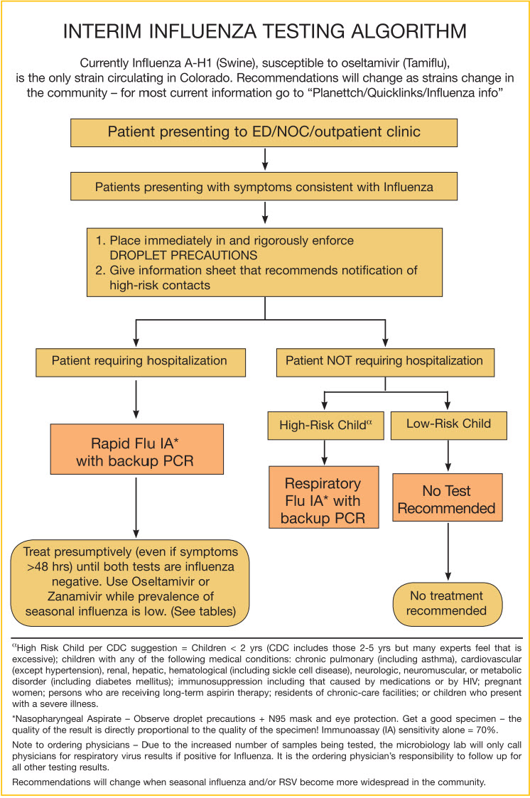
A1
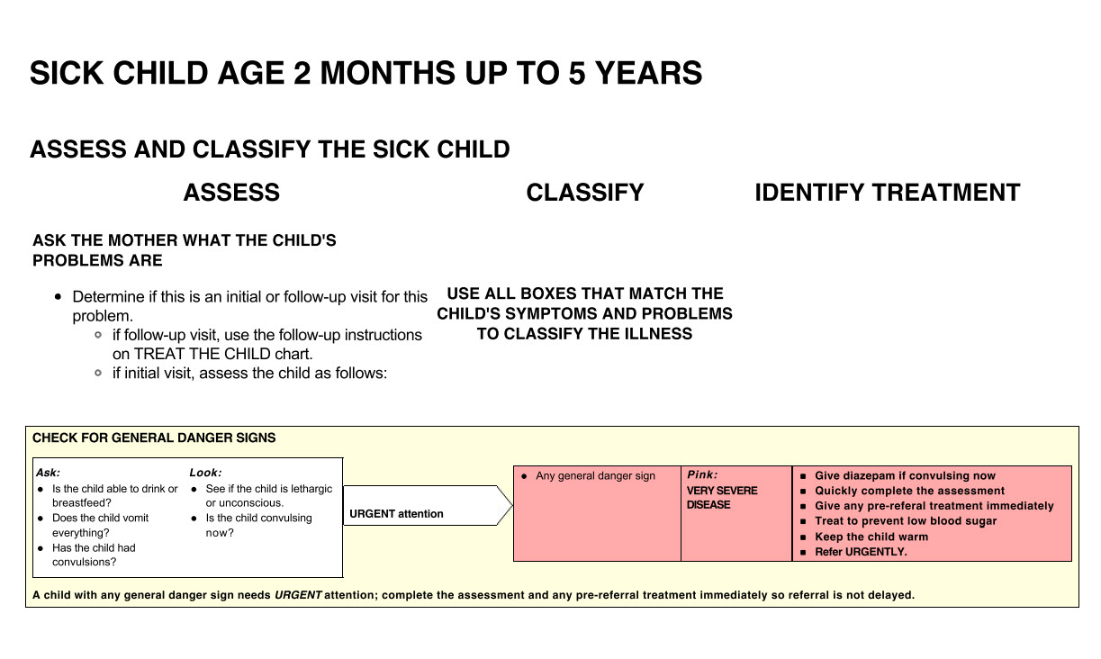
A2
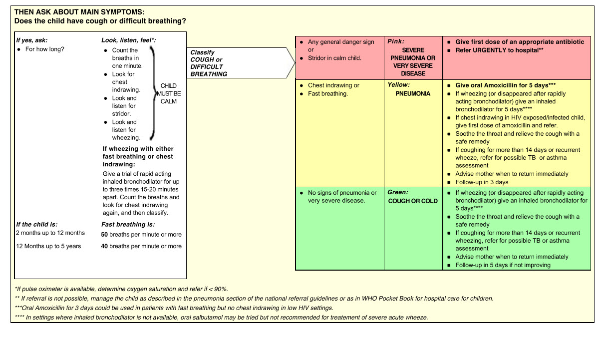
A3
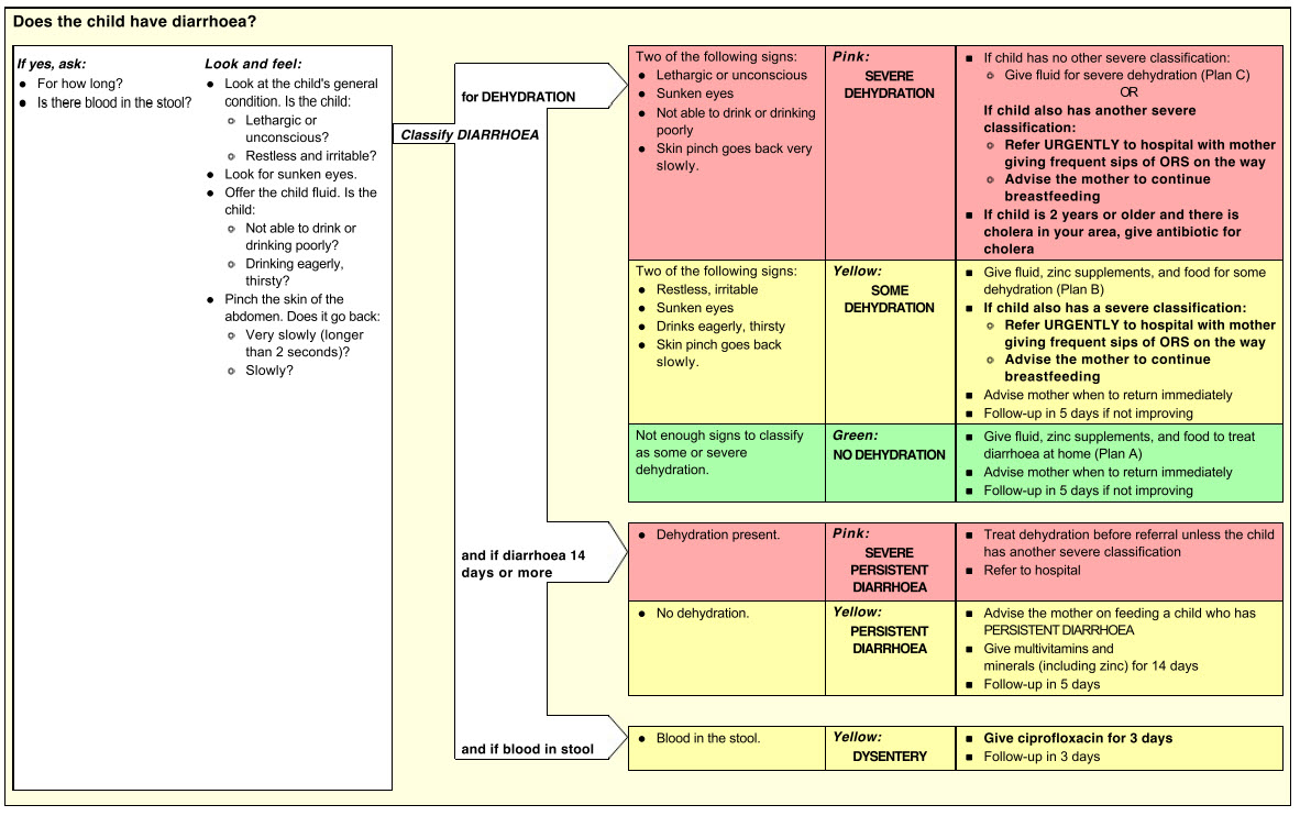
A4
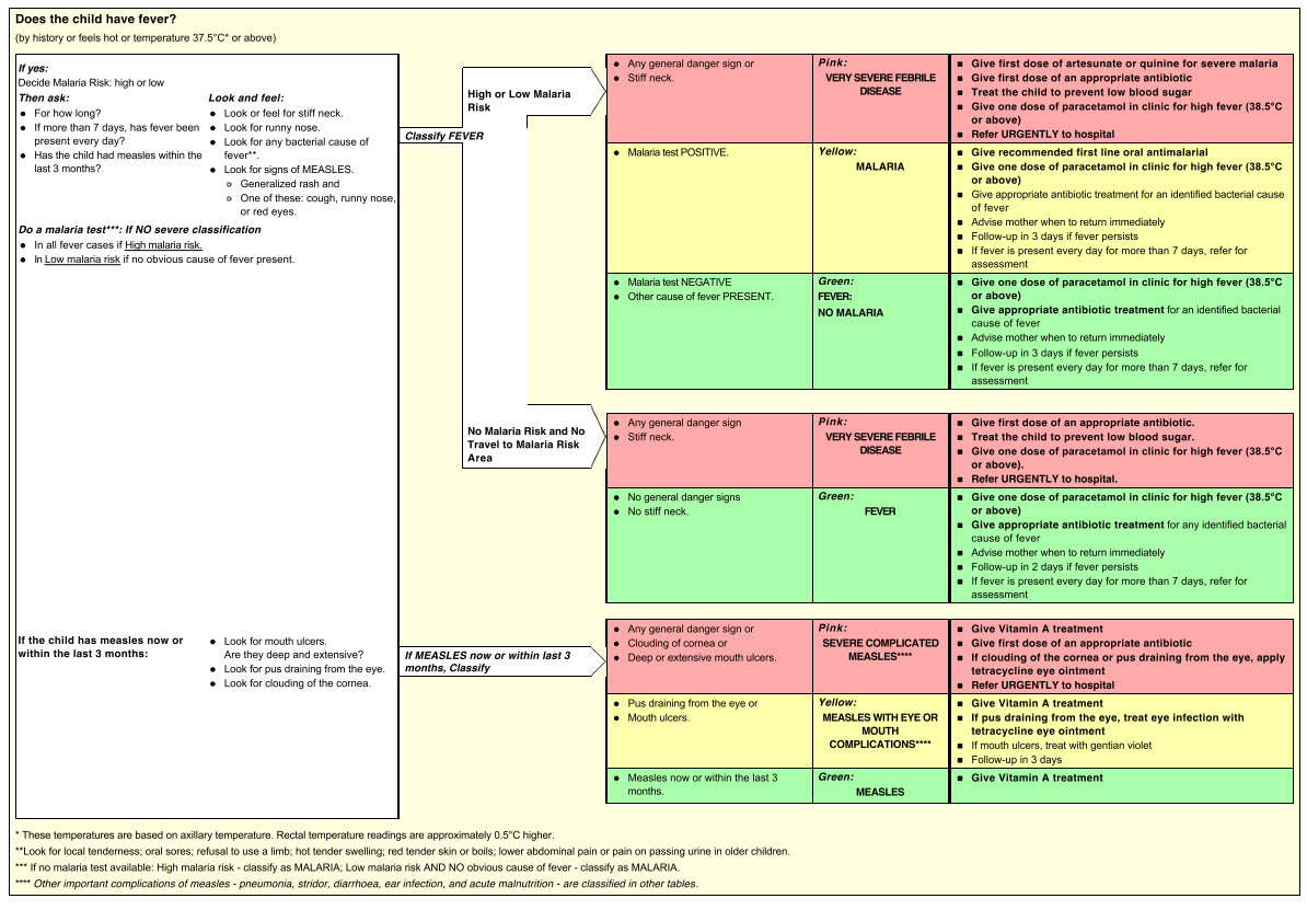
A5
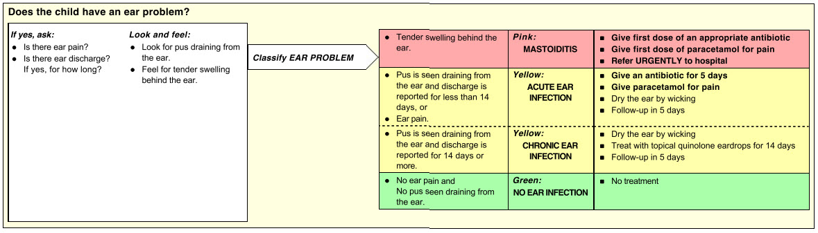
A6
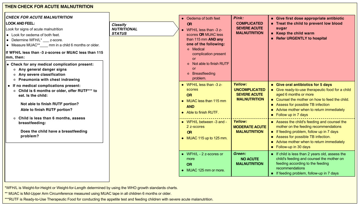
A7
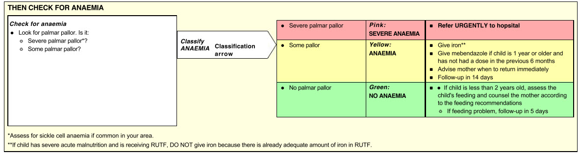
A8
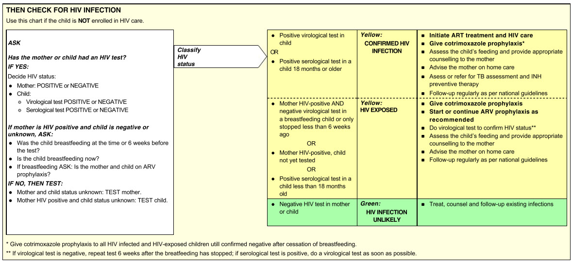
A9
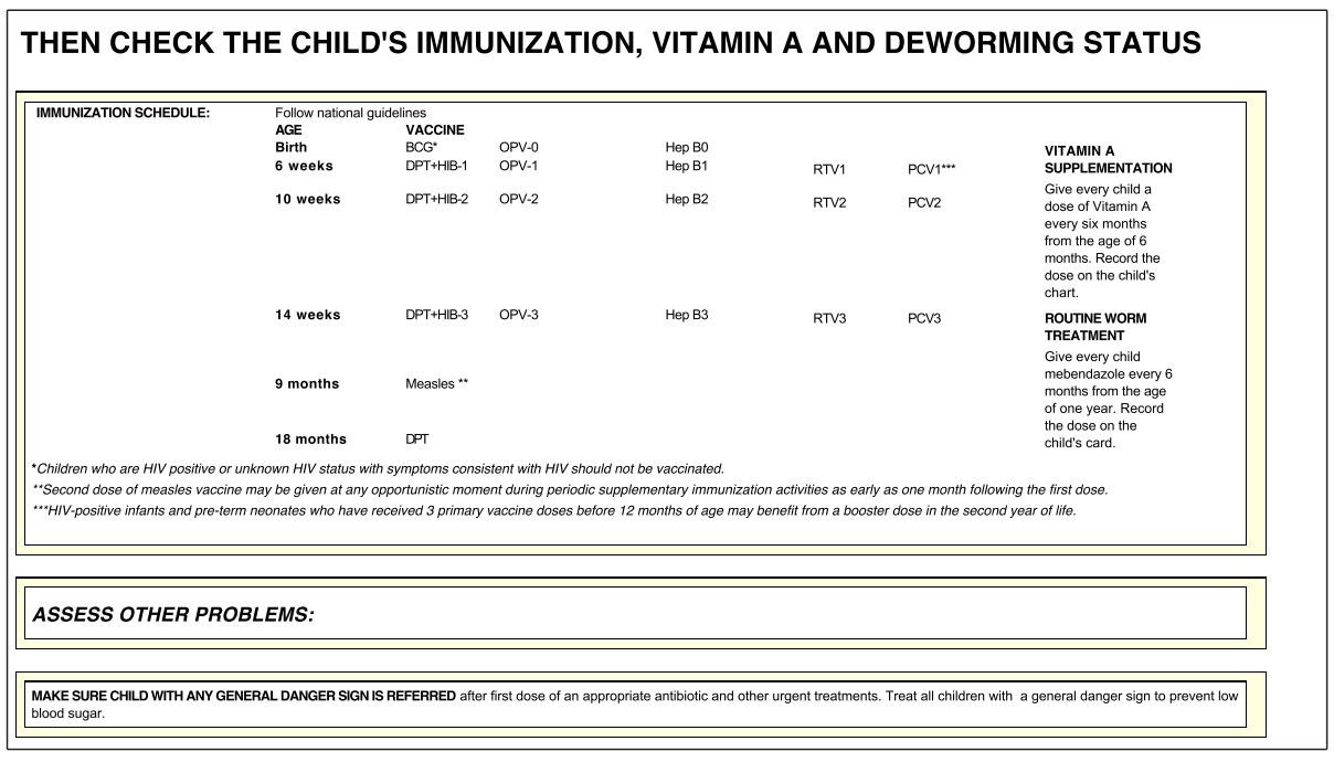
A10
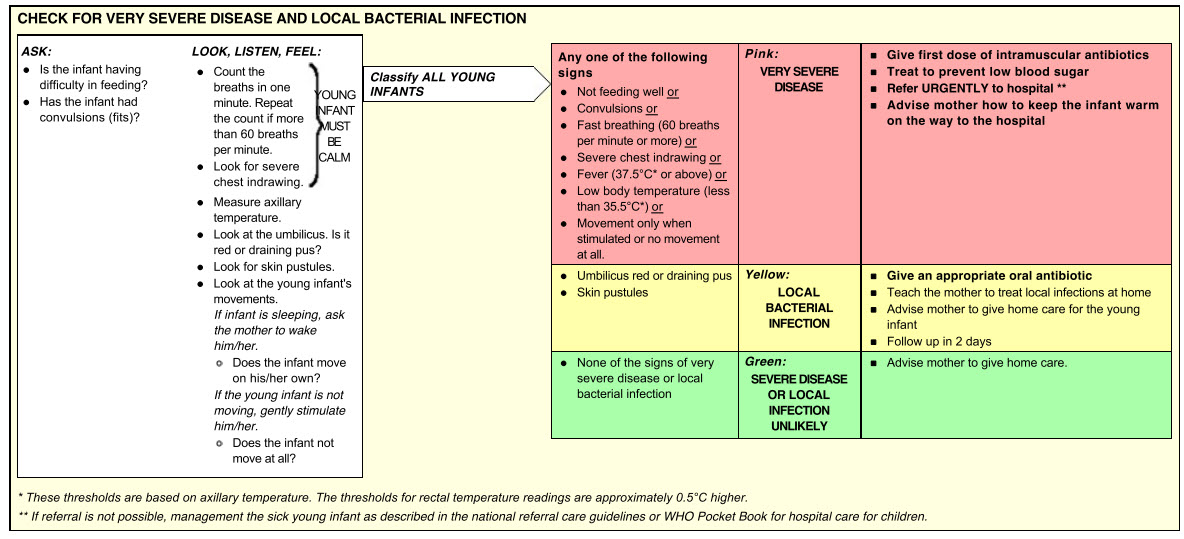
A11
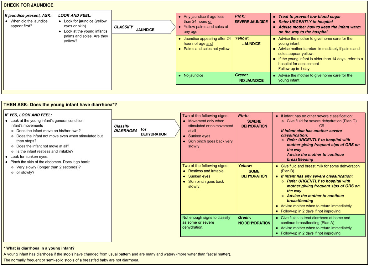
A12
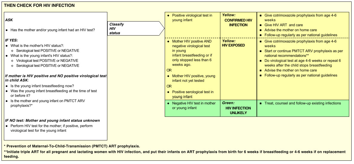
A13
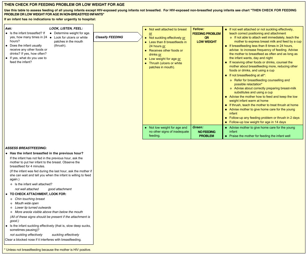
A14
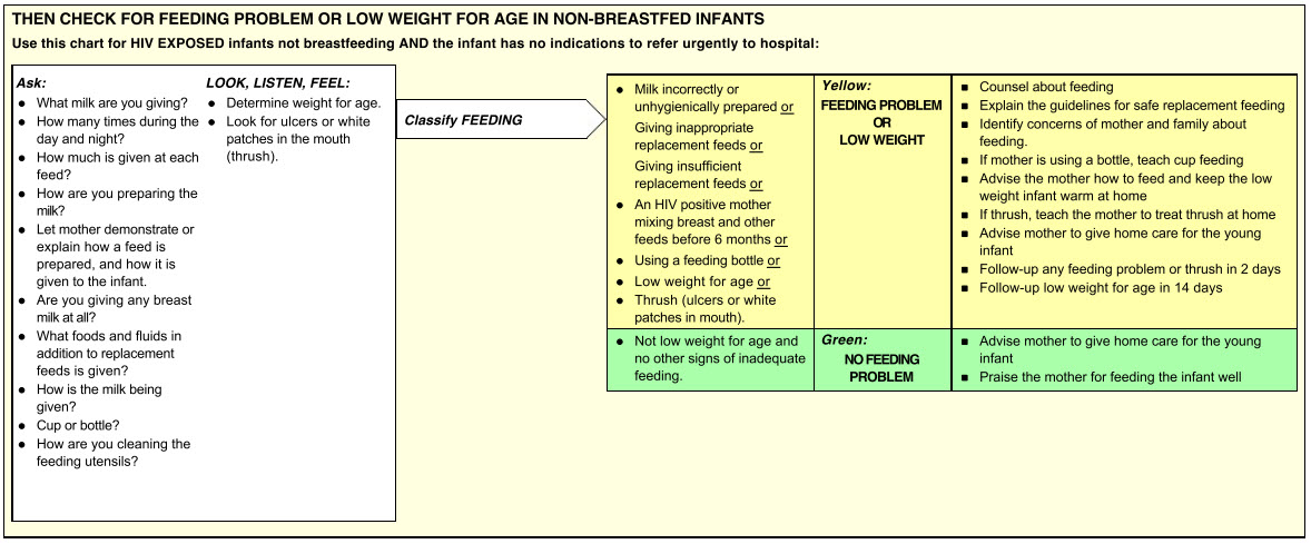
A15
A16
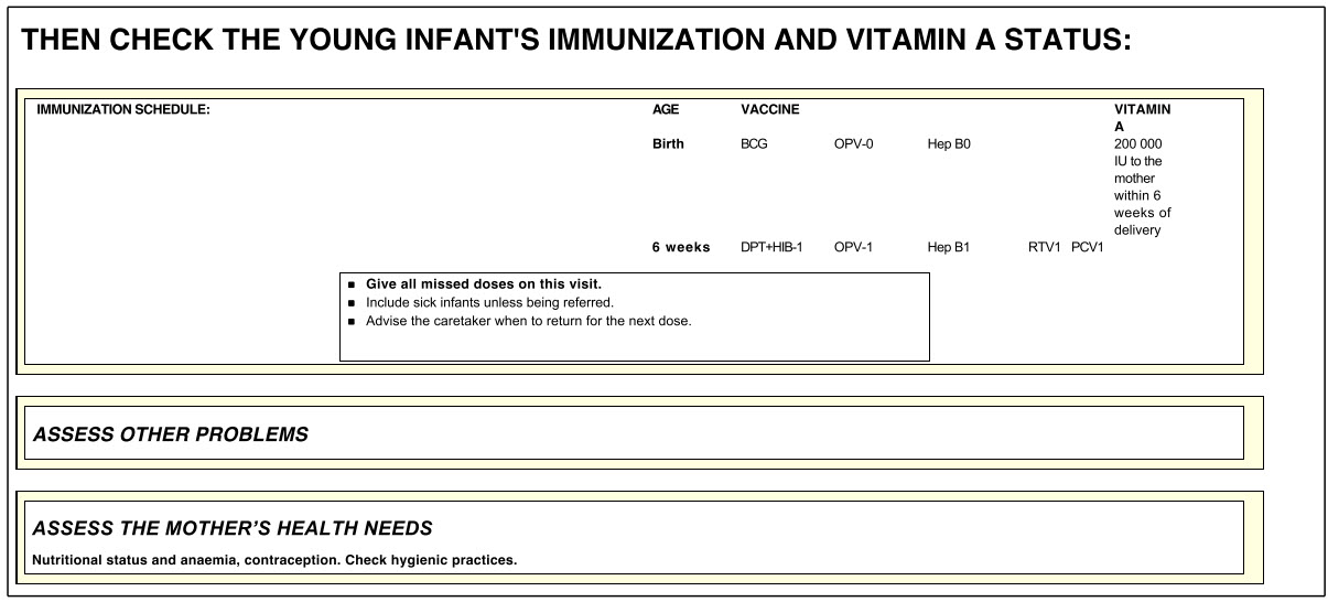
A17
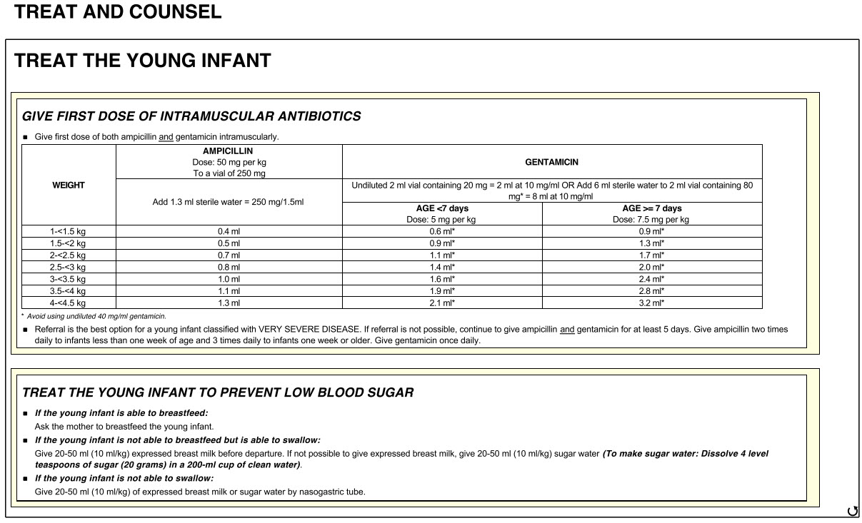
A18